The Time to Retract
Was Fischer's eighth stage a measure of rate against the tissue's capacity to escape it?
Fischer's eighth stage as a measurement of rate against host capacity, and what the modern evidence on resilience makes of it
Abstract
Oskar Fischer's 1910 monograph divides the senile plaque into eight morphological states. Seven of them are ordered in time. The eighth is not, and Fischer says so twice, in two independent ways, and gives his reason.
The eighth state is the infiltrative one: over areas sometimes larger than an immersion field, the axis cylinders and the fibrillar network have vanished and their place is taken by thread-masses without sharp borders. It looks like the end of the disease, and it is numbered last, and every subsequent reading — including every modern one — has taken it for the terminus. Fischer's own text says the opposite. He places Stage VIII outside the age series and calls it younger: the infiltrating masses take only the black impregnation, which on his own colour clock is the young colour, and he offers this as evidence that they are "Bildungen jüngeren Datums… die sich kürzere Zeit vor dem Tode gebildet haben" — formations of more recent date, laid down a shorter time before death. And he gives a mechanism for why this state looks different from the others: "das infiltrative Durchwuchern, als Ausdruck einer sehr schnellen Wucherung, bei der das nervöse Gewebe keine Zeit zur Retraktion mehr hatte." The infiltrative overgrowth is the expression of a very rapid proliferation, in which the nervous tissue no longer had time to retract.
That sentence contains a variable that the eight-stage scheme does not otherwise contain. Stages I through V are a clock: they measure elapsed time. Stage VIII is a ratio — the rate at which material is laid down, divided by the capacity of the host tissue to accommodate it. Fischer's eighth stage is not a measure of how long the disease has run. It is a measure of how badly the host was outpaced.
This paper argues three things.
First, that the reading is correct, and that it survives the checks that should have killed it. Two falsifiers were available in Fischer's own material and both fail to fire. If VIII were end-stage, the infiltrative masses should carry the old colour on his clock; they carry the young one. And if VIII were end-stage, it should track disease duration in the 1912 series the way Stages I–V do; it does not. Case 12 of 1912 — an acute delirium of a few days' standing — showed "Reichliche Aussaat kleinster Sterndrusen und massenhafte Infiltrate. Keine älteren Formen": abundant sowing of the smallest star-drusen and massive infiltrates, and no older forms at all. The infiltrative stage appears in the shortest-duration case in the series, alongside the youngest deposits, in a brain with no atrophy and no other parenchymal change.
Second, that the mechanism Fischer proposed for it is wrong, and that this does not touch the observation. He read the co-occurrence in Case 12 as evidence of an acute Schub — a wave of thread-formation laid down in the days before death, producing a delirium that could remit, with further waves producing further delirious states. Modern deposition kinetics forbid it. Longitudinal PiB-PET in 200 participants puts the transit from the threshold of amyloid positivity to the burden seen in Alzheimer's disease at 19.2 years (95% CI 16.8–22.5), with a further 12.0 years from low control levels to that threshold, in an almost linear fashion (Villemagne et al., 2013). Nothing is sown in days. What Fischer saw in Case 12 was not fresh deposition; it was, at most, a delirium in a brain that happened to carry an unusual distribution of deposit. His inference fails on the timescale. His measurement — that infiltrative material and the youngest forms occur together, and that both occur in brains the disease has not otherwise wasted — is untouched by the refutation, because it is a statement about co-occurrence and not about rate of arrival.
Third, that the salvageable core is a resilience claim, made morphologically, in 1910, on the largest human series of its era. The modern evidence that most nearly matches it is not a study of amyloid at all. It is the finding that the relationship between pathology and cognitive loss is person-specific to a degree that has no precedent in the way the disease is usually described: across 1,079 autopsied participants, Alzheimer pathology accounted for a mean of about 50% of cognitive decline but for a range of 22% to 100% at the individual level, with more than 230 distinct neuropathological combinations, none of them present in more than 6% of the cohort (Boyle et al., 2018). Capacity varies enormously between people. Fischer's eighth stage is what the low-capacity end of that distribution looks like under silver impregnation.
We then ask what the "resistance" in keine Zeit zur Retraktion could physically be, and grade three candidates: the extracellular matrix that the deposit must displace, the microglial mantle that compacts it, and the geometry of the neuropil itself. The matrix candidate is the strongest and is testable now — aggrecan is incorporated into human dense-core plaques and perineuronal nets are lost in spatial association with deposits and microgliosis (Crapser et al., 2020) — and it predicts that Fischer's infiltrative form should be found in matrix-poor territory and should be scarce where the net is intact. That prediction has never been tested, and nothing in the modern literature has ever asked it, because the stage it concerns has been read as an endpoint rather than as a rate.
We close with a graded ledger of nineteen propositions, six falsifiers, and five experiments in order. The first is a plate-level re-scoring of Fischer's own figures for colour class, which requires no tissue, no consent and no funding, and would either confirm or destroy the reading this paper is built on.
A note on what this paper does not claim. It does not claim that Fischer anticipated the modern resilience literature. He did not: he had no concept of reserve, no cognitive measurement worth the name, and his own explanation for the eighth stage was a mechanism that cannot be true. Nor does it claim that Stage VIII is rapidly progressive Alzheimer's disease; Part Four sets out four reasons why that identification fails and keeps the comparison as an analogy under grade. What is claimed is narrower: that a variable which the modern field measures indirectly and with difficulty was isolated morphologically in 1910, that it was mislaid for a century because the stage carrying it was numbered last, and that recovering it costs nothing and predicts something.
Part One — The Stage That Was Numbered Last
1. A Reading Nobody Checked
Fischer's classification table sets out eight forms of the Drusen, keyed to figures on thirteen plates. Seven of the eight are ordered. The order is defended twice — internally, by the colour of the impregnation, and externally, in 1912, against the clinical duration of the illness. It is a good piece of work, and the ordering has held up: short duration carries the younger stages, long duration the older ones, which is the 1912 antecedent of a result independently obtained a century later on forty subjects with symptom durations of four to twenty years.
The eighth form sits outside that structure, and it has been read as though it sat at the end of it.
The reading is understandable. Stage VIII is numbered eighth. It is described in language unlike anything else in the monograph — whole fields in which the axis cylinders and the fibrillar network "vollkommen geschwunden", completely vanished, replaced by the thread masses; of the nervous tissue only the ganglion cells persisting, "sehr schwer geschrumpft", severely shrunken, having lost most of their processes. Elsewhere Fischer insists that his drusen displace the tissue rather than destroying it. Here, and only here, they destroy it. A morphological state that is numbered last, looks catastrophic, and is the only one in which the tissue is genuinely wrecked, will be read as the end of the process by any reader who is not attending closely to the text.
The text says otherwise, and it says so in the two places where Fischer normally does his most careful work: the colour, and the case series.
This paper is about what follows from taking him at his word. The consequence is not antiquarian. If Stage VIII is not a terminus but a ratio, then Fischer's scheme contains a measurement of host capacity — a quantity the modern field cares about intensely, measures with difficulty, and estimates almost entirely by subtraction. And the measurement was made on 275 brains, in human tissue, with a post-mortem interval measured in minutes, by an observer who had no theory of resilience to confirm.
Three propositions organise the argument.
First, that the "younger" reading is Fischer's and is supported by two independent lines in his own material. Chapter 2 sets out the statements; Chapters 3 and 4 test them.
Second, that his mechanism for the observation is refuted and the observation is not. Chapters 5 to 8. This is the part of the paper that costs something: a reader sympathetic to Fischer must give up the Schub, which is the most vivid thing he ever wrote, and keep a duller claim underneath it.
Third, that what remains is a rate-against-capacity variable with a modern counterpart. Chapters 9 to 21, ending in three graded candidates for what the resisting material physically is.
2. What Fischer Actually Says About the Eighth Stage
Two statements, made for different reasons, in different parts of the monograph.
The first is about colour, and it is offered as evidence. Fischer's colour clock is stated at p.387: the threads impregnate black when young and brown when older. He establishes it on the internal structure of a single deposit — in the wheel-form stage the central parts are brownish to reddish while the marginal ring is always black, "daraus folgt, daß auch auf Grund des tinktoriellen Verhaltens der Randring als jüngste Bildung der Drusen erscheint" — from which it follows that on the tinctorial evidence too, the marginal ring appears as the youngest formation of the drusen. The clock is what licenses his claim that deposits grow by accretion at the rim rather than by condensation throughout.
Applied to the infiltrative masses, the same clock returns a young reading. They take only the black. Fischer draws the inference and hedges it in his own hand:
"was auch dafür sprechen könnte (ich betone hier das „könnte"), daß wir es hier mit Bildungen jüngeren Datums zu tun haben, d. h. mit Bildungen, die sich kürzere Zeit vor dem Tode gebildet haben."
("which could also speak for it — I emphasise here the could — that we are dealing here with formations of more recent date, that is, with formations that were laid down a shorter time before death.")
The parenthetical emphasis is his. It is worth noticing what kind of writer inserts it: one who knows the difference between a datum and an inference, and who is telling the reader which he is offering.
The second statement is about mechanism, and it is offered as explanation. In the passage where Fischer lists what the fungal hypothesis would explain, he includes the infiltrative mode and gives its distinguishing feature:
"das infiltrative Durchwuchern, als Ausdruck einer sehr schnellen Wucherung, bei der das nervöse Gewebe keine Zeit zur Retraktion mehr hatte"
("the infiltrative overgrowth, as the expression of a very rapid proliferation, in which the nervous tissue no longer had time to retract")
Read that against his standing position. Fischer's repeated finding is that the deposit displaces nervous tissue rather than destroying it — the halo around a druse is a retraction space, not condensed host material, and he argues the point three separate ways. Retraction is the normal response. It is what the tissue does when it has time.
Stage VIII is therefore, in Fischer's own account, the normal process running against a clock the tissue loses. The material is the same — he says so explicitly, "daß sie morphologisch aus denselben Fäserchen bestehen, die den äußeren Ring der größeren Drusen… bilden", that the infiltrating masses consist morphologically of the same fibrils that form the outer ring of the larger drusen. The mode is not a different substance. It is the same substance arriving faster than the tissue can get out of its way.
Transitional forms exist between the two modes, and Fischer found one: Fig. 40 shows a marginal ring that has attained unusual thickness, no longer bounds itself sharply, and "strahlt infiltrierend aus", radiates out infiltratingly. A druse becoming an infiltrate. That figure is the hinge of the whole reading, because it shows the two modes as endpoints of one variable rather than as two diseases.
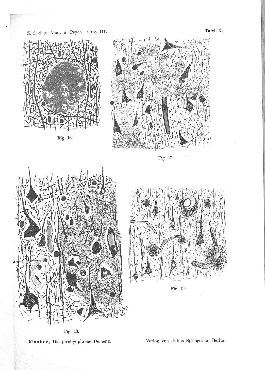
3. The First Check: The Colour Clock
If Stage VIII were the terminus of the series — the state a deposit reaches after passing through the other seven — then the infiltrative masses should carry the tinctorial signature of old material. On Fischer's clock that is brown.
They do not. They take the black.
The falsifier is clean, it was available in 1910, and it fails to fire. This is the strongest single line in the paper, and it is worth being precise about what it does and does not establish.
What it establishes. Within Fischer's own method, the infiltrative material is not old material. Whatever else Stage VIII is, it is not Stage V that has continued.
What it does not establish. That the colour clock is correct. Silver impregnation intensity depends on section depth, fixation, local pH and packing density as well as on the age of the substrate, and Fischer nowhere controls those. His defence is that the same ordering holds both within a single deposit and across deposits of different size, which is better than either alone and is still not a demonstration. If the clock is an artefact of packing density, then dense infiltrative masses would read black for reasons that have nothing to do with age — and, awkwardly for this paper, an infiltrate is exactly the kind of structure that would be densely packed.
That objection is real and this paper does not dispose of it. It is the reason the colour argument is graded supported, method-dependent in the ledger rather than established, and the reason the first experiment in Chapter 24 is a re-scoring of the plates for colour class against deposit density. If black tracks density rather than youth, the first pillar of the reading falls and the second must carry it alone.
4. The Second Check: Duration, and Case 12
The 1912 paper is where Fischer validated his ordering externally, and it is where the second falsifier lives.
His finding, at p.102, is stated plainly: "daß die Fälle mit kurzer Krankheitsdauer auch vornehmlich die jüngeren Stadien, jene mit längerer Krankheitsdauer die älteren Stadien der Drusen aufwiesen" — that the cases with short disease duration predominantly showed the younger stages of the drusen, those with longer duration the older stages. Stage tracks duration. That is the result which makes the ordering more than an aesthetic judgement, and it is the result a modern series reproduced at higher resolution a century later.
So: does Stage VIII track duration?
If it is the terminus, it must. It should appear in the longest-duration cases and be absent from the shortest. And here the 1912 series contains a case that settles the question in the wrong direction for the standard reading.
Case 12 was an acute delirium of a few days' onset. The brain showed:
"Reichliche Aussaat kleinster Sterndrusen und massenhafte Infiltrate. Keine älteren Formen."
("Abundant sowing of the smallest star-drusen and massive infiltrates. No older forms.")
Three things are in that sentence and all three matter.
The smallest star-drusen are Stage I — the youngest form in the scheme.
Massive infiltrates are Stage VIII.
No older forms at all. Not "few". None. The intermediate stages, through which a deposit would have to pass to reach a terminus, are absent.
And the brain carrying this was, in Fischer's description elsewhere in the same paper, without atrophy, with minimal glial proliferation, and with no other parenchymal change. He uses that fact for a different argument — against the idea that the drusen are the residue of decaying nervous tissue, since here is a brain full of deposit and otherwise intact — but it bears on this one too. Stage VIII appeared in a brain the disease had not wasted.
The falsifier does not fire. It does the opposite of firing: the infiltrative stage occurs in the shortest-duration case in the series, co-located with the youngest deposits, in undamaged tissue. On the terminus reading this is close to impossible. On the ratio reading it is what should be expected — a host that was outpaced does not have to have been outpaced for long.
One case is one case. The 1912 series is thirty-five brains and Fischer draws his Schub inference from two of them. This chapter does not claim that Case 12 proves the reading; it claims that Case 12 is where the standard reading had its chance to be confirmed and was not. In the ledger, the duration argument is graded supported, n small, and Chapter 24's second experiment is the one that would settle it: re-score the stage assignments across the whole 1912 series against the recorded durations, with VIII scored separately from I–V rather than folded into an ordinal scale. Fischer published the case protocols. The re-scoring is a desk exercise on printed material.
Part Two — The Mechanism He Proposed, and Why It Fails
5. The Schub
Fischer did not leave the observation as an observation. He built a clinical model on it, in spaced type, from two cases:
"daß eine ganz akute frische Sphaerotrichieaussaat zu einem akuten Delirium führt, welches in Heilung übergehen kann; die Drusen bleiben im Gehirn bestehen, nehmen allmählich die reiferen Formen an, und wenn es dann wieder zu einem Schub einer Fädchenbildung kommt, so entsteht von neuem ein deliranter Zustand."
("that a quite acute, fresh sowing of Sphaerotrichia leads to an acute delirium, which can pass into recovery; the drusen persist in the brain, gradually take on the more mature forms, and when a further Schub — a wave, a relapse — of thread-formation then occurs, a delirious state arises anew.")
It is the most attractive passage in the monograph. It converts a static morphological scheme into a disease with a course; it explains why presbyophrenic patients have fluctuating, sometimes remitting, delirious episodes rather than a smooth decline; and it makes the mature deposits a record of past attacks rather than a cause of the present one. It also has the shape of a good hypothesis: it predicts that a brain examined shortly after an acute episode will show fresh material and mature material and nothing in between, which is exactly what Case 12 shows.
It is also, on the modern evidence, not true — at least not as a statement about deposition.
6. Nineteen Years, Not Days
The kinetics are known and they are not compatible with a wave laid down in the days before death.
In a prospective cohort of 200 participants — 145 healthy controls, 36 with mild cognitive impairment and 19 with Alzheimer's disease — imaged with ¹¹C-Pittsburgh compound B every eighteen months for a mean of 3.8 years, 82% showed positive rates of amyloid accumulation, and the transit from the threshold of PiB positivity (1.5 SUVR) to the burden observed in Alzheimer's disease was estimated at 19.2 years (95% CI 16.8–22.5), progressing in an almost linear fashion at a mean of 0.043 SUVR per year. Reaching that threshold from the low levels of amyloid-negative controls took a further estimated 12.0 years (95% CI 10.1–14.9). The projected preclinical phase places the crossing of the positivity threshold about 17 years before the onset of dementia, hippocampal atrophy about 4.2 years before, and memory impairment about 3.3 years before (Villemagne et al., 2013).
Deposition is slow, protracted, and — on this evidence — nearly linear rather than episodic. Two decades is the unit. Days are not.
There is a second, structural reason the Schub cannot be rescued by arguing that PET measures bulk fibrillar load and might miss a fast local event. Fischer's model requires the mature deposits in Case 12 to be absent, and they are: "Keine älteren Formen." A brain that has been laying down deposit for two decades will contain mature forms whether or not a recent wave has occurred. The absence of intermediate stages in a brain full of Stage I and Stage VIII is therefore not evidence of a recent sowing; on the modern timescale it is evidence that something is wrong with reading the morphological series as a straightforward chronology in that brain at all.
7. What Survives
The refutation lands on the inference and not on the measurement, and it is worth separating them cleanly, because a great deal of the value of historical material is lost by readers who discard the whole of a passage when part of it fails.
Refuted: that the infiltrative masses were laid down in the days before death. Deposition does not work at that speed. Nothing in the modern record permits it.
Refuted: that a fresh sowing causes an acute delirium which can remit. Delirium in an elderly patient with cerebral pathology has a large differential — infection, metabolic derangement, drug effect, dehydration — none of which Fischer could exclude in 1912 and most of which are more probable than acute amyloidogenesis. The modern reading of Case 12 is a delirium from an ordinary cause, in a brain that happened to carry an unusual distribution of deposit.
Not refuted: that infiltrative material co-occurs with the youngest forms and without the intermediate ones. This is a statement about what was on the slide. It is a co-occurrence, not a rate, and no result about deposition kinetics touches it.
Not refuted: that Stage VIII occurred in a brain with no atrophy and no other parenchymal change. Also a statement about what was on the slide, and a more surprising one than it looks. Whatever produced those infiltrates did not produce the tissue loss that usually accompanies advanced disease.
Not refuted: that the infiltrative mode is the same material as the ordinary deposit, differing in how it relates to the surrounding tissue. Fischer's morphological identification, plus the transitional form in Fig. 40.
What is left, after the Schub is removed, is this: in some brains the deposit occupies tissue instead of displacing it, and this happens independently of how long the disease has run. That is the observation. It needs a mechanism, and Fischer's is unavailable.
8. The Cost of Keeping the Observation
It should be said plainly that this manoeuvre — the hypothesis fails, the observation survives — is the most abused move in the historical literature, and that a paper making it owes the reader a demonstration that it is not being made opportunistically.
The test is whether the surviving observation was load-bearing for the refuted hypothesis or independent of it. An observation invented to serve a dead theory usually dies with it; an observation that constrained the theory usually does not.
Here the co-occurrence in Case 12 was not what Fischer's Schub model was built to explain. The model was built to explain a clinical pattern — remitting delirious episodes in presbyophrenia — and it recruited the morphology as support. The morphological finding is prior to the model and independent of it: Stage VIII is described in 1910, two years before the Schub appears, and it is described there without any clinical claim attached, purely as a mode in which the tissue had no time to retract. The 1910 statement is the one this paper uses. The 1912 model is the one this paper discards.
That said, one genuine cost must be accepted. Without the Schub, the paper loses the only proposed explanation for why the intermediate forms are absent in Case 12. It is a real anomaly and this paper does not solve it. Three possibilities are open — sampling (the intermediate forms were present in unexamined blocks), scoring (Fischer's own admission that Stages VI–VIII "lassen sich schwieriger abschätzen", are harder to assess, applies here), or a genuine two-population structure in that brain. The third is the interesting one and it is untestable on printed plates. It is entered in the ledger as open.
Part Three — Rate Against Capacity
9. The Variable Fischer Isolated
Strip the mechanism away and read the 1910 sentence as a description of what determines which mode a deposit takes.
"eine sehr schnelle Wucherung, bei der das nervöse Gewebe keine Zeit zur Retraktion mehr hatte"
There are two terms. One is the rate at which material accumulates. The other is the time the tissue needs to retract — which is another way of saying the tissue's capacity to accommodate an intruding mass without being destroyed by it. Fischer's eighth stage occurs when the first exceeds the second.
This is a different kind of quantity from anything else in his scheme, and the difference is worth stating formally.
Stages I–V are a clock. They report elapsed time since the deposit began. A brain full of Stage IV has been depositing longer than a brain full of Stage II. This is why the ordering could be validated against disease duration, and why it was.
Stage VIII is a ratio. It reports the relation between two rates. A brain full of Stage VIII has been outpaced — and it may have been outpaced quickly or slowly, recently or long ago. The ratio carries no information about elapsed time, which is precisely why Stage VIII does not track duration and should not have been expected to.
Numbering the ratio as though it were the eighth position on the clock is the error, and it is a notational error rather than an observational one. Fischer put it in the table because the table was his list of forms, and he then told the reader, twice, that this one was not in the series. Everyone since has read the table and not the caveats.
There is a modern analogue for exactly this mistake, and naming it may help. Braak staging orders the topographic spread of neurofibrillary pathology, from the brainstem outward and from the third decade of life (Braak & Del Tredici, 2011); it is a clock. The phases of amyloid deposition order the spread of the deposit through the brain (Thal et al., 2002); also a clock. Neither scheme has a slot for how well the individual brain was tolerating what it had — and the field's repeated discovery that stage predicts cognition only loosely is, in part, the discovery that a clock is being asked to do the work of a ratio.
10. Two Brains, the Same Burden, Different Outcomes
The observation that pathology and clinical state come apart is old, and the case that established it in the modern literature was a subgroup with preserved mental status and numerous neocortical plaques (Katzman et al., 1988): a substantial fraction of people at high pathological stage are not demented at death, and a substantial fraction of dementia in the community is not attributable to Alzheimer neuropathological change of any grade.
Fischer had the same observation, in a cruder form and from the other end. His Case 12 is a brain crowded with deposit and otherwise entirely intact. His 1912 discussion of latency and threshold is an attempt to say what such a brain means:
"eine Art latenter Gehirnveränderung… die erst dann zu klinischen Zeichen geführt hätte, wenn sie zu stärkerer Entwicklung gekommen wäre"
a kind of latent brain change, which would have led to clinical signs only when it had come to stronger development; and, in the same passage, that the process "erstens bereits eine Zeit lang bestehen muß, bevor die klinischen Symptome sich entwickeln, und zweitens ein bestimmter Grad der anatomischen Laesion notwendig ist" — must first have existed for some time before clinical symptoms develop, and second, that a certain degree of the anatomical lesion is necessary.
Time, and a threshold. He is describing a preclinical phase and a tolerance limit, in 1912, from thirty-five non-demented elderly brains of which only two carried deposits.
What he lacked was any way to measure the second term. The threshold is asserted; nothing in his material lets him say whether it differs between people, or by how much. That is the gap the modern evidence fills, and it fills it dramatically.
11. How Much Capacity Varies
The most useful number for this argument comes from a study that was not about amyloid.
In 1,079 participants from two longitudinal clinical–pathologic cohorts, each with two or more cognitive evaluations before death and a full neuropathological examination for nine pathologies, mixed pathology was the rule rather than the exception: 94% had at least one, 78% at least two, 58% at least three, 35% at least four. More than 230 distinct neuropathological combinations were observed, each present in fewer than 6% of the cohort. Alzheimer pathology was the most frequent at 65% but occurred in isolation in only 9%. And the crucial figure: although Alzheimer pathology accounted for a mean of about 50% of observed cognitive loss, the proportion it accounted for at the individual level ranged from 22% to 100% (Boyle et al., 2018).
Read that as a statement about capacity rather than about comorbidity. Two people may carry the same amyloid burden and be at opposite ends of a fourfold range in how much of their cognitive loss that burden explains. Whatever determines the difference is not the deposit. It is the host.
This is the modern form of the term Fischer could name and not measure. And it makes his eighth stage interesting in a specific way: if capacity varies that much between individuals, there should be a morphological signature of low capacity, and it should be visible in tissue. Fischer proposed one. Nobody has looked for it, because the stage he proposed it in has been read as an endpoint.
12. Why "Stage" Was the Wrong Word
A short chapter, because the point is simple and consequential.
A stage is a position in a sequence. Calling the infiltrative mode a stage licenses three inferences, all of which are false on Fischer's own account:
- that a deposit passes through it (his transitional Fig. 40 shows entry into the mode, not passage through it to something else);
- that it comes after the others (he says twice it is younger);
- that it can be ordinally scored with the others in a single scale (which is what every subsequent use of his table has done, and which mixes a clock and a ratio in one column).
The third is the one with practical consequences. Any re-scoring of Fischer's material — including the re-scoring proposed in Chapter 24 — must score VIII on a separate axis, or the ratio will be averaged into the clock and disappear. This is not a subtlety. It is the mechanism by which the finding was lost in the first place.
Part Four — The Modern Fast Form
13. Rapidly Progressive Alzheimer's Disease
If there is a modern entity in which deposition outpaces accommodation, the obvious candidate is the rapidly progressive form of the disease.
It exists, it is neuropathologically confirmed, and it is defined clinically rather than morphologically. In a multicentre retrospective series drawn from France, Germany, Japan and Spain, 89 neuropathologically confirmed cases of rapidly progressive Alzheimer's disease — cases initially classified as prion disease on the strength of their clinical phenotype — had a median survival of 10 months. Cerebrospinal fluid biomarkers were abnormal but within the range expected for classic Alzheimer's disease, except that the 14-3-3 proteins were detectable in 42%. The genetic profile is the striking part: APOE and PRNP codon 129 genotype distributions paralleled healthy controls, and ε4 homozygosity was absent (Schmidt et al., 2012).
14. The Genotype That Is Missing
That last finding deserves emphasis, because it is the one that makes the comparison worth drawing at all.
APOE ε4 is the principal common risk allele for Alzheimer's disease and it acts, in large part, on burden: ε4 carriers deposit more amyloid, earlier, and ε4 is a dose-dependent risk factor for cerebral amyloid angiopathy — one copy raising the odds of moderate or severe angiopathy 2.9-fold and two copies 13.1-fold in systematically graded post-mortem material (Greenberg et al., 1995). If the fast clinical form were simply the high-burden form running harder, ε4 homozygotes should be over-represented in it.
They are absent from it.
Whatever makes the disease run fast is therefore, on this evidence, not the same thing as what makes it deposit heavily. That is the modern shape of Fischer's distinction: rate of deposition and the outcome of the encounter are separable variables, and the second is not a monotone function of the first.
15. What a Rate Disease Should Look Like at Autopsy
The prediction is straightforward and, as far as we can establish, untested.
If the infiltrative mode indexes the ratio rather than the burden, then in rapidly progressive Alzheimer's disease one should find:
- deposit morphology skewed toward the infiltrative, ill-bounded, non-retracted form;
- not necessarily a higher total plaque count than in classic disease;
- less well-formed peri-deposit retraction space (Fischer's halo) per deposit;
- and, on the reasoning of Part Five, less intact perineuronal matrix in the affected territory.
None of these has been looked for. The neuropathological description of rapidly progressive cases has concentrated on excluding prion disease and on establishing that the Alzheimer changes are present and adequate — which is a different question from whether the deposits are shaped differently.
16. Where the Analogy Breaks
Four reasons the identification of Stage VIII with rapidly progressive Alzheimer's disease must not be made, and why this paper keeps it as an analogy under grade.
The clinical entity is defined by survival, not by morphology. Rapidly progressive Alzheimer's disease is a phenotype selected for speed of clinical decline. Fischer's Stage VIII is selected for the appearance of the deposit. There is no reason in advance why the two selections should pick out the same brains, and Case 12 — a delirium with an intact brain — is a warning that they may not.
Fischer's cases were presbyophrenic and not necessarily what would now be diagnosed. The 1907–1912 material is a clinical population defined by a category that no longer exists. Mapping it onto a modern diagnostic entity is exactly the kind of transfer that generates false continuities.
Speed of clinical decline has causes that are nothing to do with the deposit. Comorbid pathology, delirium, systemic illness and hospitalisation all accelerate measured decline; the association between hospitalisation rate and faster cognitive decline persists after controlling for seven neuropathological markers, and is stronger in those with more tangle pathology and neocortical Lewy bodies (James et al., 2019). A fast course is not evidence of a fast deposit.
The absence of ε4 homozygosity in the rapidly progressive series may be a selection artefact. The cases were referred because they were mistaken for prion disease. Whatever determines that referral pattern could plausibly correlate with genotype through the clinical phenotype rather than through the biology.
The comparison is entered in the ledger as suggestive, unmatched — the two literatures make compatible claims about the separability of rate and burden, and no study has examined the same brains for both.
Part Five — What Resists
17. Retraction Is a Physical Act
The whole argument turns on a word Fischer used without defining it. Retraktion. The tissue retracts, when it has time.
It is worth taking the word literally, because he did. His position on the halo — the clear space around a mature druse — is that it is a space and not a condensation: not host tissue compressed into a rim, but host tissue that has withdrawn, leaving a gap. He argues it three ways and the argument is one of the better pieces of reasoning in the monograph.
If the halo is a retraction space, then retraction is something the neuropil does, over time, in response to a slowly enlarging mass. It is the mechanical accommodation of an intruder. And a tissue's ability to perform it will depend on properties that are, in principle, measurable: how much the local matrix can be remodelled, how fast, and by whom.
That reframes the question of Part Five. "What resists?" is really: what must be remodelled for a deposit to be accommodated rather than to destroy? Three candidates, graded.
18. The First Candidate: The Matrix
The strongest candidate is the extracellular matrix, and specifically the condensed lattice that surrounds a subset of cortical neurons.
The relevant modern finding is that this matrix is not a bystander to deposition. Aggrecan — the principal lectican of the condensed matrix — is incorporated into human dense-core plaques; in the 5xFAD model, perineuronal net abnormality and loss occur in spatial association with thioflavin-S-positive dense-core deposits across the course of deposition, and in association with IBA1-positive microgliosis; and microglia engulf net material (Crapser et al., 2020). The causal arm of that work is murine and the human arm is descriptive, which is how it is graded here and in the source.
Two things follow for this paper.
The matrix is on the path. A deposit enlarging in cortex is enlarging in a space that contains this material, and the material ends up inside the deposit. Whatever else is happening, the lattice is being remodelled or overrun.
The remodelling is enzymatic and therefore rate-limited. A lattice that must be cut before the tissue can give way is exactly the kind of structure that would produce Fischer's dichotomy: give it time and the tissue accommodates and retracts; exceed the rate at which it can be cut, and the deposit occupies the space instead.
This candidate makes the paper's sharpest prediction, in Chapter 23: the infiltrative mode should be found preferentially in matrix-poor territory, and should be rare where the net is intact. Cortical layers differ substantially in net density, and Fischer recorded a laminar gradient in deposit density — richest in the upper layers, decreasing downward. Whether the mode also has a laminar gradient he does not say, and his plates may be able to answer it.
Grade: inference, testable. The link from "aggrecan is in the plaque" to "matrix capacity determines deposit mode" is not established by any experiment. It is a hypothesis with an available test.
19. The Second Candidate: The Mantle
The second candidate is the cell that arrives at the deposit and organises it.
Microglia form a barrier around dense-core deposits that compacts the amyloid and limits the protofibrillar Aβ42 at its margin — the mantle is a containment structure, not merely a reaction to one (Condello et al., 2015). Its adequacy is genotype-dependent in human material: TREM2 R47H shifts deposit morphology and the associated neuritic injury at unchanged total burden (Yuan et al., 2016). And the local features of individual deposits — dystrophic neurites, CD68-positive microglial activation, GFAP-positive astrocytes — rise with symptom duration while plaque burden does not, and while microglial number does not move at all (Serrano-Pozo et al., 2016).
That last dissociation is the important one for this argument. What changes over the clinical course is not how many microglia there are but what they are doing. A containment function that can be adequate or inadequate at constant cell number is a capacity term in exactly the sense this paper needs.
Against the candidate: Fischer could not see it. Bielschowsky silver does not demonstrate microglia, so nothing in his material can bear on whether the mantle is what failed in his infiltrative fields. Any argument from his plates to the mantle is an argument from absence, and this paper does not make one.
Grade: plausible, invisible to the source material. It is a good candidate for the biology and a poor one for the historical claim.
20. The Third Candidate: The Geometry
The third candidate is that nothing is being remodelled at all, and the difference between the modes is architectural.
The neuropil is not homogeneous. A deposit growing in a region of dense, parallel, myelinated fibre traffic occupies a different mechanical environment from one growing in a felt of unmyelinated terminals. Fischer's own distribution data is at least consistent with an architectural term: the drusen are richest in the upper cortical layers, decrease downward, and are absent from the white matter altogether — and the white matter is the most mechanically ordered compartment of the three.
This candidate has the advantage of requiring no biology, and the disadvantage of predicting almost nothing. If the mode is fixed by local architecture, it should be highly stereotyped by region and should not vary between individuals with the same regional sampling. That is a real prediction and it is the one Chapter 23 uses to separate this candidate from the first.
Grade: weak, but not excluded. It is entered chiefly because it is the null hypothesis for the matrix account, and a paper that proposes a resistance term should say what it would mean for the term to be trivial.
21. What Is Not Being Claimed
Three disclaimers, because the argument of Part Five is the most speculative in the paper and the easiest to over-read.
No claim that the matrix is the resistance. Three candidates are offered, one is preferred, and the preference rests on a testable prediction rather than on evidence in hand.
No claim that Fischer had any of this. He had a word — Retraktion — and an observation that it sometimes fails. He had no concept of the extracellular matrix as an organised structure, and the condensed perineuronal lattice was described by a contemporary of his whose work he does not cite.
No claim that the resistance is a single thing. The three candidates are not mutually exclusive and the honest expectation is that accommodation is multi-factorial, in which case Fischer's dichotomy is a threshold on a composite and the search for "the" resisting material is misconceived. That possibility is entered in the ledger, and it is the one under which this paper's central prediction would fail while its central observation remained true.
Part Six — Consequences
22. A Graded Ledger
Three levels are used. Established — directly evidenced, in human material or by convergent human and animal data. Supported — good evidence with a stated dependency. Inference — consistent with the evidence, not directly demonstrated. Two further labels are used where they are more honest than a grade: open, and beyond the evidence.
Three rows deserve comment.
Row 15 is the paper, and it is graded beyond the evidence. That is deliberate. The proposition that Fischer's eighth stage is a morphological readout of host capacity is a proposal, not a finding, and every experiment in Chapter 24 exists to move it up or delete it. A reader who takes away only the ledger should take away that the strongest claim in this paper is the least supported one in it.
Row 5 carries the reading and has a live objection. The colour argument depends on Fischer's clock being a clock. If black tracks density, an infiltrate reads black for trivial reasons and the first pillar collapses. Experiment 1 tests exactly this, on printed plates, and it should be run before anything else in this paper is relied upon.
Row 12 is an anomaly the paper does not solve. Removing the Schub removes the only available explanation for the missing intermediate forms in Case 12, and nothing replaces it. It is left open rather than filled, because the three candidate explanations — sampling, scoring, a genuine two-population structure — cannot be separated on printed material.
Each claim below carries a tier and, where it is not settled, the observation that would settle it. 7 claims · 1 not yet settled
Strong — Amyloid deposition takes about two decades, not days
Villemagne 2013, n = 200, longitudinal PiB
What would settle it. — not in serious dispute
Strong — Fischer's Schub model of acute sowing is therefore false
9
What would settle it. —
Strong — Host capacity to tolerate Alzheimer pathology varies severalfold between individuals
Boyle 2018, n = 1,079; 22–100% person-specific range
What would settle it. —
Established (supported, method-dependent) — Therefore the infiltrative material is not old material
3 + 4
What would settle it. Evidence that impregnation intensity tracks packing density rather than age
Established (supported, n small) — Stage VIII does not track disease duration as Stages I–V do
1912 Case 12; the 1912 duration finding
What would settle it. A full re-scoring of the 1912 series showing VIII concentrated in long-duration cases
Established (supported) — Rate of decline and burden of deposit are separable
Schmidt 2012 (ε4 homozygosity absent in rpAD); Greenberg 1995 (ε4 dose raises burden)
What would settle it. Replication of the rpAD genotype finding in a prospectively ascertained series
Weak (beyond the evidence) — Stage VIII is that signature
The paper's central proposal
What would settle it. The experiments of Chapter 24
23. Predictions and Falsifiers
Six, each stated so that its failure counts against the paper.
P1. In any re-scoring of Fischer's plates, infiltrative material will fall in the black colour class and will not show the brown of the mature deposits. Falsified by: brown infiltrative material at appreciable frequency.
P2. In a re-scoring of the 1912 case protocols with VIII scored on a separate axis, the presence of infiltrates will be uncorrelated, or negatively correlated, with recorded disease duration, while Stages I–V remain positively correlated. Falsified by: infiltrates concentrated in the longest-duration cases.
P3. In modern human material, deposits in territory of high perineuronal net density will more often show a well-formed retraction space than deposits in net-poor territory, at matched deposit diameter. Falsified by: no association, or the opposite association.
P4. The infiltrative, ill-bounded deposit morphology will be over-represented in rapidly progressive Alzheimer's disease relative to classic disease at matched total burden. Falsified by: equal or lower representation at matched burden. The burden-matching is essential; without it the prediction is trivial.
P5. Deposit mode will vary between individuals more than local architecture alone predicts — that is, two individuals sampled in the same cortical region and layer will differ in mode more than two regions within one individual. Falsified by: mode being fixed by region, which would support the architectural null of Chapter 20 and remove the need for a host-capacity term.
P6. APOE ε4 dose will predict burden more strongly than it predicts mode. Falsified by: ε4 dose predicting mode as strongly as burden, which would collapse the rate/burden distinction this paper depends on.
24. Five Experiments, in Order
1. Re-score the plates for colour class against deposit density. No tissue, no consent, no funding. The thirteen plates of the 1910 monograph are public domain and the figures are keyed to stages in Fischer's own table. Score each figure for colour class and for packing density, blind to stage assignment, and test whether colour is predicted by density. This decides row 5 of the ledger, and it can be done in a week. It is first because it can destroy the paper.
2. Re-score the 1912 series with VIII on a separate axis. Fischer published the case protocols with recorded disease durations. Extract stage assignments and durations, score the infiltrative finding as a separate binary rather than as the eighth point of an ordinal scale, and test P2. Also a desk exercise on printed material.
3. Retraction space against matrix density in modern human cortex. Dual-label a cortical series for dense-core deposits and for perineuronal net material, measure per-deposit halo area at matched deposit diameter, and stratify by local net density. Tests P3, which is the discriminating prediction of the matrix candidate. Requires ordinary autopsy material and no new methodology.
4. Deposit mode in rapidly progressive Alzheimer's disease. The rapidly progressive series exist and are neuropathologically confirmed. Score deposit mode and total burden in rapidly progressive and duration-matched classic cases. Tests P4. The obstacle is access to banked material rather than method.
5. Mode variance decomposition. In a series sampled at fixed cortical coordinates across many individuals, partition the variance in deposit mode into between-region and between-individual components. Tests P5, and thereby separates the host-capacity account from the architectural null. Largest of the five and the only one requiring a purpose-built cohort.
25. Limitations
The central proposition is unfalsifiable in Fischer's material and untested in anyone else's. Experiments 1 and 2 test the historical reading; 3, 4 and 5 test the biological proposal; and until at least one of the latter is run, row 15 stays where it is.
The colour clock may be an artefact, and if it is, the paper loses a pillar. This is stated in Chapter 3, graded in row 5, and tested first in Chapter 24. It is the most likely way for the argument to fail.
One case is not a series. The duration argument rests on Case 12 plus the general 1912 finding. Fischer himself drew the Schub from two cases and was wrong; a paper that criticises him for that cannot rest its own weight on one.
Fischer's own scoring of the late stages is admittedly weak. "Die drei letztgenannten Stadien lassen sich schwieriger abschätzen" — the last three stages are harder to assess. He says it, and it applies with full force to the stage this paper is about. Nothing here strengthens his scoring; the argument is built on his statements about Stage VIII rather than on his assignments of it.
The presbyophrenic population is not a modern diagnostic category. Every inference from his clinical material to a modern entity crosses a nosological gap that cannot be closed retrospectively.
Whether any of Fischer's slides survive is not established here. Experiment 3 is designed for modern material precisely so that it does not depend on the answer.
26. Conclusion — The Ratio in the Table
Fischer's table has eight rows and seven of them are a clock. The eighth is a ratio, and he said so twice — once in the language of colour, once in the language of mechanism — and then put it in the table anyway, because the table was a list of forms and that is what he had.
The century that followed read the table and not the caveats. The eighth row was taken for the end of the disease because it was numbered last and looked like ruin, and a variable that no other part of the scheme contained went missing inside an ordinal scale.
What went missing is worth recovering. The modern field knows that host capacity varies severalfold between individuals, that this variation dominates the relation between pathology and clinical state, and that it is measured almost entirely by subtraction — by what the pathology fails to explain. Fischer proposed a positive morphological readout of the same quantity: not how much deposit there is, and not how long it has been there, but whether the tissue was given time to get out of its way.
He was wrong about why. Deposition takes two decades and nothing is sown in the days before death, so the Schub goes, and with it the most vivid paragraph he ever wrote. The observation underneath it does not go: in some brains the deposit occupies tissue instead of displacing it, and this happens in brains the disease has not otherwise wasted, and it happens independently of how long the disease has run.
Whether that is the signature of a low-capacity host is a question with five experiments attached, the first of which needs a copy of the plates and a week. It has been available since 1910.
Sources and Reproductions
The plates of the 1910 monograph (Tafel VII–XIX, published by Julius Springer, Berlin) are in the public domain and are reproduced from the original printing.
All German quotations are taken from the original papers and are located by journal page in the text. Translations are the author's, with the German given alongside wherever the reading turns on it.
No modern micrographs are reproduced. The figure this paper would most want — deposit mode scored against perineuronal net density in human cortex — does not exist, which is the substance of Experiment 3.
References
Boyle PA, Yu L, Wilson RS, Leurgans SE, Schneider JA, Bennett DA. Person-specific contribution of neuropathologies to cognitive loss in old age. Annals of Neurology. 2018;83(1):74–83. PMID 29244218. DOI 10.1002/ana.25123
Braak H, Del Tredici K. The pathological process underlying Alzheimer's disease in individuals under thirty. Acta Neuropathologica. 2011;121(2):171–181. PMID 21170538.
Condello C, Yuan P, Schain A, Grutzendler J. Microglia constitute a barrier that prevents neurotoxic protofibrillar Aβ42 hotspots around plaques. Nature Communications. 2015;6:6176. PMID 25630253. DOI 10.1038/ncomms7176
Crapser JD, Spangenberg EE, Barahona RA, Arreola MA, Hohsfield LA, Green KN. Microglia facilitate loss of perineuronal nets in the Alzheimer's disease brain. EBioMedicine. 2020;58:102919. DOI 10.1016/j.ebiom.2020.102919
Fischer O. Miliare Nekrosen mit drusigen Wucherungen der Neurofibrillen, eine regelmässige Veränderung der Hirnrinde bei seniler Demenz. Monatsschrift für Psychiatrie und Neurologie. 1907;22:361–372. Find this paper
Fischer O. Die presbyophrene Demenz, deren anatomische Grundlage und klinische Abgrenzung. Zeitschrift für die gesamte Neurologie und Psychiatrie. 1910;3:371–471. (Mit 13 Tafeln.) Find this paper
Fischer O. Ein weiterer Beitrag zur Klinik und Pathologie der presbyophrenen Demenz. Zeitschrift für die gesamte Neurologie und Psychiatrie. 1912;12:99–135. (Mit 1 Tafel.) Find this paper
Greenberg SM, Rebeck GW, Vonsattel JP, Gomez-Isla T, Hyman BT. Apolipoprotein E ε4 and cerebral hemorrhage associated with amyloid angiopathy. Annals of Neurology. 1995;38(2):254–259. PMID 7654074. DOI 10.1002/ana.410380219
James BD, Wilson RS, Capuano AW, Boyle PA, Shah RC, Lamar M, Ely EW, Bennett DA, Schneider JA. Hospitalization, Alzheimer's disease and related neuropathologies, and cognitive decline. Annals of Neurology. 2019;86(6):844–852. PMID 31614018. DOI 10.1002/ana.25621
Katzman R, Terry R, DeTeresa R, Brown T, Davies P, Fuld P, Renbing X, Peck A. Clinical, pathological, and neurochemical changes in dementia: a subgroup with preserved mental status and numerous neocortical plaques. Annals of Neurology. 1988;23(2):138–144. PMID 2897823.
Schmidt C, Haïk S, Satoh K, Rábano A, Martinez-Martin P, Roeber S, Brandel JP, Calero-Lara M, de Pedro-Cuesta J, Laplanche JL, Hauw JJ, Kretzschmar H, Zerr I. Rapidly progressive Alzheimer's disease: a multicenter update. Journal of Alzheimer's Disease. 2012;30(4):751–756. PMID 22460329. DOI 10.3233/JAD-2012-120007
Serrano-Pozo A, Betensky RA, Frosch MP, Hyman BT. Plaque-associated local toxicity increases over the clinical course of Alzheimer disease. The American Journal of Pathology. 2016;186(2):375–384. PMID 26687817. DOI 10.1016/j.ajpath.2015.10.010
Thal DR, Rüb U, Orantes M, Braak H. Phases of Aβ-deposition in the human brain and its relevance for the development of AD. Neurology. 2002;58(12):1791–1800. PMID 12084879.
Villemagne VL, Burnham S, Bourgeat P, Brown B, Ellis KA, Salvado O, Szoeke C, Macaulay SL, Martins R, Maruff P, Ames D, Rowe CC, Masters CL. Amyloid β deposition, neurodegeneration, and cognitive decline in sporadic Alzheimer's disease: a prospective cohort study. The Lancet Neurology. 2013;12(4):357–367. PMID 23477989. DOI 10.1016/S1474-4422(13)70044-9
Yuan P, Condello C, Keene CD, Wang Y, Bird TD, Paul SM, Luo W, Colonna M, Baddeley D, Grutzendler J. TREM2 haplodeficiency in mice and humans impairs the microglia barrier function leading to decreased amyloid compaction and severe axonal dystrophy. Neuron. 2016;90(4):724–739. PMID 27196974. DOI 10.1016/j.neuron.2016.05.003
Genes named on this page: ApoE, apolipoprotein E; ACAN (aggrecan), aggrecan; TREM2; PRNP; GFAP; CD68; AIF1 (Iba1), IBA1.