The Geode and the Flower
What happened to Fischer's observations after him?
Oskar Fischer's staged deposit, 1907–1912, read against the intracellular accounts of Gunnar Gouras and Ralph Nixon
Abstract
In 1910 Oskar Fischer set out the two available explanations for the senile plaque and rejected both. The first was that the deposit is the residue of nervous tissue falling apart in place. The second — which he stated in a single sentence, in words that describe the amyloid cascade with uncomfortable accuracy — was that it is a soluble species produced at one site, carried elsewhere, precipitated at a distance, and assembled crystal-like into a fibrillar mass. He declined the first because he found a brain crowded with deposits and otherwise intact. He declined the second because the orderly progression of forms he had documented seemed to him too structured for a precipitate. Having refused both, he concluded that the material was "dem Nervensystem morphologisch und chemisch ganz Fremdes" — morphologically and chemically wholly foreign to the nervous system — and spent four independent experimental lines testing whether it was an organism. All four came back negative.
The twentieth century adopted the hypothesis Fischer had written down and turned down. This paper argues that the third option, the one his own data pointed at and his vocabulary could not name, has since been reconstructed by two laboratories working eighty years later and largely independently of one another: that the deposit is neither a precipitate from a distance nor the generic decay of tissue, but the exported or spilled interior of one particular cell. Gunnar Gouras established that the nerve cell manufactures amyloid-β on the organelles it uses to run its synapses, and that the pathogenic pool is the fraction retained rather than released. Ralph Nixon established that the disposal network fails at its terminal acidification step, that the swollen neurite is packed almost to the exclusion of everything else with vesicles of that failed pathway, and that a neuron distended with de-acidified autolysosomes can rupture and leave behind an object with the appearance of a cored plaque.
Read against those two accounts, six of Fischer's observations change status. Four of them he recorded without explanation and they now have one; two of them constrain the modern claim rather than confirming it.
The clods are lipid. At his seventh stage Fischer treated the dissolving deposit with Marchi's method and found the granules within it blacken — a lipid reaction, in a lesion the modern era classifies as a protein aggregate. He concluded the clods were breakdown products of the threads themselves rather than of cells, because he could not find enough transitional forms between a cell and a clod. His dichotomy had no third term. The third term is a membrane-bounded organelle, and the predominant organelles of the Alzheimer dystrophic neurite are autophagosomes, multivesicular bodies, multilamellar bodies and autolysosomes.
The nuclei are the disagreement, and it is quantifiable. Fischer reported nuclei and nuclear detritus inside the larger deposits, of provenance he could not determine, and called such occurrences "immerhin eine Seltenheit" — nonetheless a rarity. Ninety-one years later a human immunohistochemical series reported a nuclear remnant at the dense core of many plaques and read each plaque as the end product of a single neuronal lysis. These are two direct human observations of the same structure, in opposite directions, on the question a modern evaluation of the intracellular account marks as unanswered: what fraction of the human plaque burden formed from within. We show that both figures are uncorrected two-dimensional section counts of a three-dimensional coincidence, that the sampling geometry alone predicts a severalfold discrepancy in that direction, and that the correction is arithmetic rather than conceptual. Corrected, Fischer's figure bounds the intracellular fraction from below rather than from above: read as a rate rather than as an adjective, his Seltenheit is evidence for a substantial intracellular contribution and not against one. It is an unanalysed measurement of the quantity in dispute, and it is the oldest one in existence.
The neurite rule is a statement about a living cell. Fischer's central quantitative finding is that the club-shaped axonal swellings cluster around his fifth and fourth stages, are rarer at the third, and are absent at both ends of the series: "nie in der Nähe der Stadien I und II, ebenso auch nie um Stadium VIII." The relation of injury to deposit is an inverted U, not a ramp. A curve of that shape cannot be produced by a deposit corroding its surroundings; it requires the club to be something a surviving neurite does, with a window that opens when there is enough stimulus and closes when there is no longer a process in a condition to respond. That is the shape both modern programmes predict and neither has stated, and it remains untested in human tissue.
We also record where the legacy does not hold. Fischer's sixth stage, the fur-like destruction of the vessel wall, is cerebral amyloid angiopathy, and it is the compartment in which the extracellular route is strongest and the intracellular route has least to say — the peptide that defines the modern disease was purified from it. The diffuse, non-fibrillar deposit that constitutes the bulk of the amyloid load in the unimpaired elderly was invisible to his stain and is explained fully by neither account. And his conclusion of foreignness was an error of exactly the kind that mattered: material can be host-derived and still show no transition to host tissue, if it arrives packaged inside a membrane and is released as a unit. The membrane is the term his dichotomy lacked, and its absence is why the man who wrote the amyloid hypothesis down in 1910 was unable to accept it.
Finally, the paper returns Alzheimer's objection to its original form. In 1911 Alzheimer argued against Fischer that the drusen displace nervous structures more than they destroy them, that dementia occurs where drusen are few, and that degeneration occurs in regions where drusen are absent — and concluded that the deposits are not the cause of the disease but an accompaniment to it. Fischer conceded the first premise. The argument has been restated in every decade since without its authorship being noticed. Its three limbs are answered, not evaded, by an account in which the plaque is partly a record of a death that occurred inside a cell: a gravestone displaces without destroying, because the destruction happened before the marker existed; the disposal lesion is general while the deposit-leaving morphology is conditional on a cargo only some cells make; and the number of markers is a poor index of the number of deaths for reasons that are geometric as well as biological.
We close with a graded ledger of twenty-three propositions, seven falsifiers, and six experiments in rank order. The first is a three-dimensional nuclear-remnant count in human neocortex, which would settle in one study a question that has been open since 1910 and which no laboratory has yet performed.
Part One — The Question Fischer Asked
1. Why This Comparison
Oskar Fischer is cited for priority and read for almost nothing else. The standard sentence — that he described the plaque in 1907 in a larger series than Alzheimer's single case, and was then eclipsed — is true, and it has become a substitute for engaging with what he actually established. The consequence is that a body of human morphological data assembled on 275 brains, with artefact controls that would not embarrass a modern paper and a post-mortem interval measured in minutes, sits unused while the field argues about questions those data bear on directly.
This paper takes one of those questions. It is the question of where the plaque comes from, and it is live. Two laboratories have spent, between them, sixty-four years building an account in which the deposit originates inside a neuron rather than condensing outside one. Gunnar Gouras's programme has established that the peptide is manufactured on synaptic machinery and that a fraction is retained rather than exported. Ralph Nixon's has established that the terminal step of the cell's disposal system fails, that the resulting undegraded cargo fills the neurite and the perikaryon, and that a cell so distended can rupture and leave a deposit behind. Both programmes are frequently described as inverting the standard picture. Both are open to the objection that a finished plaque carries no record of how it formed.
Fischer looked at the same object with a silver impregnation and drew what he saw. He was not testing the intracellular hypothesis, because it did not exist; he was testing two others and rejecting both. That is precisely what makes his record useful. Observations made under a hypothesis tend to confirm it. Observations recorded by a careful observer who could not account for them are a different kind of evidence, and there are several of them in the 1910 monograph — a lipid reaction in a protein deposit, nuclei whose provenance he could not establish, a core empty of axons and a rim crowded with them, an injury curve with two zeroes.
Three propositions organise what follows.
First, that the modern intracellular accounts answer questions Fischer posed and could not close. He asked, explicitly and in print, whether the deposit was local decay or distant precipitation. He answered no to both. There was a third possibility his vocabulary could not express, and the two programmes examined here have supplied it.
Second, that Fischer's data constrain those accounts as well as supporting them. His observation on the frequency of nuclei inside deposits is the oldest human measurement bearing on the fraction of plaques formed from within, and it points the opposite way from the only modern measurement of the same thing. Neither is usable as reported. Both become usable when the sampling geometry is corrected, and the correction is the paper's principal technical contribution.
Third, that his central quantitative finding has never been tested. The inverted-U relation between deposit stage and neuritic injury is the sharpest claim in his scheme, it discriminates between two classes of mechanism, and no modern series has measured it.
A note on what this paper is not. It is not an argument that Fischer anticipated the modern accounts. He did not; he rejected the hypothesis that turned out to be closest to right, for a reason that was intelligent and wrong, and Chapter 24 sets out the error precisely. Nor is it an argument from priority. That a claim was made in 1910 is not evidence that it is true, and where his observations disagree with better instruments the better instruments win. What is being claimed is narrower and, we think, more useful: that a large, carefully controlled, human morphological dataset exists which was gathered before any of the modern hypotheses were formulated, that it speaks to a question the modern hypotheses cannot settle from their own material, and that nobody has asked it.
2. The Disjunction of 1910: Two Origins, Both Rejected
The relevant passage is on page 389 of the 1910 monograph, and it is short. Fischer has finished describing the eight forms of the deposit and turns to the question of what produces them.
He disposes of the first possibility on the evidence of the smallest deposits. If the drusen were simple destruction-masses of nervous tissue, they would have to be found in direct connection with tissue elements undergoing destruction. They are not. The smallest forms "liegen unvermittelt in dem sonst nicht weiter destruierten Nervengewebe" — lie without mediation in nervous tissue otherwise not further destroyed. He had made the same point in the description of Stage I: "Zugrunde gegangene Elemente oder in Destruktion befindliche Gewebsbestandteile sieht man in der nächsten Nähe der Sternchen nie" — one never sees perished elements or tissue components in destruction in the immediate vicinity of the little stars.
He returns to it in 1912 with a case rather than a generalisation. Case 12 was an acute delirium of a few days' onset. The brain showed "Reichliche Aussaat kleinster Sterndrusen und massenhafte Infiltrate. Keine älteren Formen" — an abundant sowing of the smallest star-drusen and massive infiltrates, no older forms — in a brain with no atrophy, minimal glial proliferation and no other parenchymal change. A brain full of fresh deposit and nothing else wrong with it, Fischer writes, "beweist wohl wieder die Unwahrscheinlichkeit eines derartigen Ursprunges" — again demonstrates the improbability of such an origin. Whatever the deposit is, it is not the wreckage of neurons falling apart.
Then comes the second possibility, and it is worth reading slowly:
"dagegen könnten es etwa solche Abbauprodukte sein, welche aus einer ursprünglich gelösten Form fern vom Orte ihrer Entstehung niedergeschlagen werden und krystallähnlich zu Drusen sich formen. Gewisse morphologische Ähnlichkeiten sind zwar hier, aber die verschiedene Form, Tinktion und Anordnung in den verschieden großen Drusen spricht doch nicht dafür."
("On the other hand they might be breakdown products which are precipitated from an originally dissolved form, far from the site of their origin, and assemble crystal-like into drusen. Certain morphological similarities are indeed present here, but the different form, tinction and arrangement in the differently sized drusen do not speak for it.")
A soluble species, generated at one place, transported, deposited elsewhere, nucleating into an ordered fibrillar assembly. Every element of the amyloid model is in that sentence, written seventy-four years before the peptide was purified. Fischer weighed it and turned it down, and his reason is stated: the variety of form, tint and arrangement across deposits of different size did not look to him like the behaviour of a precipitate.
He also names the transport route the hypothesis would require, and rules it out on anatomical grounds: "es gehört zur Regel, daß Abbauprodukte den Weg der perivaskulären Lymphräume nehmen, aber dann füllen die Massen auch das Lumen der Spalträume aus; die Drusen sitzen aber trotz ihrer perivaskulären Anordnung in dem Gewebe selbst." It is the rule that breakdown products take the way of the perivascular lymph spaces, but then the masses also fill the lumen of those spaces; the drusen, despite their perivascular arrangement, sit in the tissue itself. The route he invokes and dismisses is the one now held to carry the peptide out of the parenchyma, and whose failure is held to produce the vascular deposits of his own sixth stage.
Having rejected both, he draws the conclusion that follows from a false dichotomy:
"daß wir in diesen Drusen etwas dem Nervensystem morphologisch und chemisch ganz Fremdes vor uns haben."
Something morphologically and chemically wholly foreign to the nervous system. The rest of the 1910 discussion is an attempt to find out what foreign thing it might be, and it is far more rigorous than the usual account of it. He states the case for a filamentous organism at length and honestly — build-up from threads, colour and thickness varying with age, apparently active growth with consequent displacement of the tissue, infiltrative overgrowth as the expression of very rapid proliferation, close attachment to vessels and growth through the vessel wall — and then kills it with a single observation: "Aber! — wo bleibt dann die entzündliche Reaktion?" Fungal growths that provoke no inflammatory reaction are unknown. He backed the negative with bacterial stains, with systematic histology of most organs of the body in several cases, with culture "in der mannigfaltigsten Variation", and with complement-fixation serology on drusen-bearing brains. All negative. In 1912 he restated, in his own defence, that he had raised the Streptothrix comparison "ohne dieselben je als tatsächliche Bakterien hingestellt zu haben" — without ever having presented them as actual bacteria.
So the position at the end of the 1910 monograph is: not local decay, not distant precipitation, not an organism, and chemically foreign. That is not a theory. It is the shape of a hole, and the hole has a definite outline. What would fill it is a material that is generated by the host, that arrives at its site of deposition already assembled rather than by precipitation, that shows no continuity with the surrounding neuropil, and that is chemically unlike the fibrillar architecture of the tissue around it. Every clause of that description is satisfied by the contents of a ruptured organelle.
3. The Limits of One Silver Stain
Fischer opens the 1910 monograph on technique, and the reason he gives is not modesty. His lesion is a lesion of one stain, and he says so as an explanation of the historical record:
"Die Veränderung im Gehirn, um die es sich handelt, läßt sich am besten mit der Methode von Bielschowsky darstellen. Mit den meisten der bisher üblichen und histologischen Färbemethoden werden die in Frage kommenden Elemente gar nicht gefärbt oder sind so wenig deutlich, daß sie sehr leicht übersehen werden können; dies ist auch der Grund, weswegen diese häufige Veränderung erst so spät bekannt wurde."
A structure invisible to the reagents in general use is a structure the field walks past. He also understood the corollary — that a lesion demonstrable by exactly one impregnation invites the charge that the impregnation is manufacturing it — and answered it with controls. He reports the same formations across a panel of chemically unrelated methods. He notes that the large drusen are visible in unfixed, unstained fresh frozen section, and states correctly that this observation "schließt eine jede Diskussion über Kunstprodukte aus". And he engineered the post-mortem interval down to minutes: "in dem größten Teil der Fälle wurde sofort nach dem Tode mittels einer Lumbalpunktionskanüle Formol in den Duralsack injiziert und auf diese Weise das Rückenmark und Gehirn in situ anfixiert." Formol injected into the dural sac through a lumbar-puncture cannula immediately after death, fixing brain and cord in place.
That last control matters more for this paper than for any other use of Fischer, and it deserves emphasis. Autophagic and endolysosomal compartments are the organelles most sensitive to post-mortem delay; it is why the definitive quantification of what fills the dystrophic neurite had to be done on antemortem cortical biopsy tissue rather than on autopsy material (Nixon et al., 2005). Fischer's material was fixed within minutes of death, in situ, in the majority of his cases. Whatever else is true of his preparations, they are not degraded in the direction that would erase the structures at issue here.
Against that stand four specific blindnesses, and being exact about them is what allows his observations to be used at all.
He could not see the non-fibrillar deposit. Bielschowsky is a fibrillar impregnation. It demonstrates the drusen and it is relatively blind to the loose, diffuse, non-fibrillar deposits that constitute the bulk of the amyloid load in cognitively unimpaired elderly brains and which a modern immunohistochemical series finds readily. His Stage I is therefore not the first deposit but the first fibrillar deposit. There is a stage zero in front of his series, invisible to him, and on modern evidence it is the most numerous form of all. Chapter 23 takes up what this costs.
He could not see microglia. Bielschowsky silver does not demonstrate them. The mantle of plaque-associated microglia that compacts the deposit and holds protofibrillar species off the neuropil (Condello et al., 2015) was not merely misread by Fischer; it was absent from his preparations. Everything he concluded about the deposit, he concluded without any knowledge of the cell that builds it. Chapter 12 shows that this statement needs one qualification, because he did see a different glial reaction, and saw it stage-dependently.
He could not identify the parent cell of a process. His clubs are identified as axis-cylinder swellings by their staining and fibrillar structure. He had no means of typing the neuron they belonged to, no transmitter markers, and no way to distinguish an axonal from a dendritic profile with confidence in every case.
He had no chemistry beyond solubility and staining reaction. His 1912 solvent series is a genuine piece of analytical work — acids, alkalis, concentrated salt solutions, permanganate oxidation with sulphurous-acid reduction, ether-alcohol, xylol, acetone — and it delivered the correct answer that the threads are "eine eiweißartige Substanz", a protein-like substance, against Marinesco's view that they were lipoid. But a stain reaction is not a molecular identification, and Chapter 8 turns on one such reaction whose meaning Fischer could not have known.
The methodological asymmetry that results is worth stating plainly, because it governs how his evidence should be weighted throughout this paper. Fischer's positive observations are strong: what he reports seeing, in in-situ-fixed human tissue, with cross-method controls, is very likely to have been there. His negative observations are weak in exactly the places where his stain was blind, and the blindnesses are known and enumerable. A finding of "never" about axon-related structures carries weight. A finding of "never" about cells does not.
4. Geode, Flower, Plaque
Fischer named the lesion twice, and both names describe an interior.
The 1907 name was drusige Nekrose, and the adjective is mineralogical. A Druse in German is a geode — a rock cavity lined with inward-pointing crystals — and the 1910 text makes the derivation explicit: the next-larger forms "auf Grund ihres regelmäßigen Baues noch eine größere Ähnlichkeit mit Krystalldrusen besitzen", on account of their regular construction possess a still greater similarity to crystal druses. The smallest he calls "kleinste Krystallsternchen", tiny crystal stars, about two micrometres across. He complained in 1912 that French authors had misread the word as drüsig, glandular, and foisted on him the notion of a nécrose glandulaire: "Was man sich wohl darunter verstellen mag?" — what one is supposed to imagine by that, he wonders. The complaint is not pedantry. "Glandular" imports a secretory, acinar, host-tissue architecture; the geode imports a foreign mineral growth built from the inside outward in a cavity, which is what he meant and what his colour data supported.
The second name is the one he formally proposed, in the classification section:
"Zu diesem Zwecke würde ich den Namen: Sphaerotrichia cerebri multiplex vorschlagen, einen Ausdruck, der nichts anderes anzeigen soll, als daß es sich um eine in meist kugeliger Form auftretende Fädchenbildung handelt."
Sphaira, sphere; thrix, thread. A thread-formation appearing in a mostly spherical form. Again: architecture, and specifically internal architecture.
Neither name survived. The field kept plaque — a word for a patch on a surface, silent about internal organisation — and after Divry's 1927 demonstration of birefringence appended amyloid, a word about the staining chemistry of the centre. The pair that replaced Fischer's pair describes the lesion's location and its core chemistry. His described its construction.
It is worth noticing what happened next, because the naming and the science moved together. A vocabulary that treats the lesion as a patch on a surface makes the natural unit of measurement the number of patches. A vocabulary that treats it as a spherical thread-formation with an interior makes the natural unit its state of construction. The twentieth century took the first and produced the plaque count, which is the measure that famously fails to predict dementia — a summary statistic over a population that Fischer had already published evidence was heterogeneous in exactly the dimension determining the outcome.
Which brings us to the third name, and to the reason this paper carries the title it does. In 2022 Nixon's laboratory described the terminal morphology of a neuron that has lost the ability to acidify its autolysosomes: very large numbers of amyloid-positive compartments packed into petal-like blebs bulging from the perikaryal membrane and arranged around the cell in a corona, after which the cell ruptures and leaves behind something with the appearance of a classical cored neuritic plaque. They named it PANTHOS, from the Greek — a poisonous anthos, a poisonous flower (Lee et al., 2022).
A geode and a flower. One is a mineral cavity lined with crystals growing inward; the other is a corona of petals arranged around a centre. Both are metaphors of internal construction, chosen a hundred and twelve years apart by observers looking at the same object, and both were chosen in preference to a word about surfaces. That is not evidence of anything. But it is a reasonable indication that two people who looked hard at the structure and its interior reached for the same class of description, and that the word which displaced the first of them was chosen by people who were looking at something else.
Part Two — The Two Modern Answers
5. The Synapse Makes It Where It Works
Gunnar Gouras began publishing on amyloid in 1998 and has continued for twenty-eight years. The programme is not a hypothesis defended over time but a sustained line of laboratory work with one object of study: where the peptide is made inside a nerve cell, what it does there, and what that has to do with the failure of synapses. Only the parts that bear on the origin of the deposit are set out here.
Location determines product. In 1999, with Greenfield and Xu in Greengard's laboratory, the group established by three independent approaches — imaging in intact neurons, density-gradient separation of compartments, and cell-free reconstitution — that Aβ40 is generated in the trans-Golgi network and packaged for export, while a population of Aβ42 is generated and retained in the endoplasmic reticulum in an insoluble state (Greenfield et al., 1999). Amyloid is not one substance made at one site; it is a family of products sorted by geography, some destined to leave the cell and some not.
The first cut happens on synaptic machinery. The two enzymes competing for the initial cleavage of the precursor protein — BACE1, which begins the amyloidogenic route, and ADAM10, which pre-empts it — are both strongly enriched on synaptic vesicles isolated from rat brain, with ADAM10 activity and the intermediate fragments detectable in the same fraction; of the complex performing the second cut, only one component is enriched, and active enzyme co-localises with the synaptic-vesicle marker only sparsely (Lundgren et al., 2015). The synapse is not merely exposed to amyloid. It manufactures the precursor fragments locally, on the organelles it uses to transmit.
The peptide accumulates inside human neurons before the deposit exists. In 2000 the group reported that Aβ42 accumulates within neurons in the regions vulnerable to Alzheimer's disease, in a distribution appearing to precede both plaques and tangles (Gouras et al., 2000). Two years later, by electron microscopy, that intraneuronal peptide was localised to multivesicular bodies inside synaptic terminals, associated with abnormal synaptic structure before plaque pathology was present (Takahashi et al., 2002); in 2004 it was seen assembling into small soluble clusters within those same processes (Takahashi et al., 2004). Independently, Cataldo, Nixon and colleagues had shown that swollen early sorting compartments precede amyloid deposition in sporadic disease and in Down syndrome (Cataldo et al., 2000). Two lines converged on the same compartment from different directions.
The pathology is retention, not overproduction. This is the least-cited turn in the programme and the most consequential for the present argument. In diseased neurons the amount of amyloid secreted falls with time in culture rather than rising, normal neurons show no such decline, and the ability of synaptic activity to increase secretion while reducing the internal pool becomes impaired; the mechanism identified is loss of surface neprilysin, the enzyme that activity normally recruits to degrade the peptide at the membrane (Tampellini et al., 2011). The diseased synapse is not making more. It is disposing of less, and failing to export what it has made.
Fibrils form inside individual synaptic compartments. Three-dimensional reconstruction from confocal imaging using thioflavin S — a dye that binds β-sheet structure rather than a sequence — showed fibrillar amyloid within individual synaptic compartments, associated with abnormal morphology and in places appearing to pierce the cell membrane (Capetillo-Zarate et al., 2011). Interfering with the packaging machinery that carries cargo into multivesicular bodies traps amyloid inside the neuron and enlarges the compartment (Edgar et al., 2015; Willén et al., 2017a).
And the structural change precedes the plaque, measured without antibodies. The standing objection to all of this has been that the antibodies used cannot reliably distinguish the peptide from the precursor it was cut from — an objection whose most candid statements have come from Gouras himself, including the finding that the standard immunoassay underestimates amyloid once it has assembled (Stenh et al., 2005) and the observation that detergent used in tissue processing can remove the intraneuronal pool altogether (Gouras et al., 2012). The answer was to drop the antibody. Synchrotron-based infrared micro-spectroscopy reports β-sheet conformation as a physical signature; applied to brain tissue it showed that the structural states of both peptide and precursor are altered before any plaque forms, and that focal aggregates preceding plaque formation localise to synaptic terminals (Klementieva et al., 2017).
Gouras has drawn from this an explicit origin claim, the inside-out amyloid hypothesis: that plaques form principally from the rupture of amyloid-laden neurons and processes, so that the deposit is the residue of a cellular failure rather than its cause (Gouras, 2014). The claim is coherent and it is supported in model systems. It is also the part of the programme with no quantitative human support, and one test of a related prediction in a knock-in animal expressing humanised sequence at normal levels went the other way: cerebral deposition preceded rather than followed the fall in fluid Aβ42/Aβ40 ratios (Andersson et al., 2023). We take the mechanism as established and the origin claim as a live hypothesis, which is how the evidence supports it.
6. The Cell Cannot Digest What It Made
Ralph Nixon's programme began in 1990 with an observation nobody could place. Cataldo and Nixon reported that senile plaques in Alzheimer brain contain lysosomal proteases, and that the proteases are enzymatically active (Cataldo & Nixon, 1990). Cathepsin D and related hydrolases, normally held behind a lysosomal membrane, were present in the extracellular deposit and retained catalytic capacity there.
It is worth pausing on how odd this was in 1990. The plaque had been understood since the sequencing of the peptide as an extracellular aggregate of a secreted product. On that reading there is no reason for the interior of a lysosome to be in it. Lysosomal contents in the neuropil imply either exocytosis of lysosomal material on a substantial scale, or the rupture of cells. Neither implication was pursued. The finding was filed as a curiosity about plaque composition.
The programme that grew from it holds the following, in the compressed form relevant here.
The endosome swells first. Neurons in sporadic Alzheimer's disease show markedly enlarged early endosomes with increased hydrolase delivery, present at early neuropathological stages (Cataldo et al., 1997); the enlargement precedes amyloid deposition, is present in Down syndrome in some neurons before birth, is accentuated by APOE ε4, and was not found in the comparison diseases examined (Cataldo et al., 2000). Among structural abnormalities in this disease it has the best claim to be the first.
The driver is the fragment, not the peptide. β-Secretase cleavage leaves a ninety-nine-residue carboxy-terminal fragment in the membrane, βCTF or C99. Elevated βCTF on the early endosome recruits the Rab5 effector APPL1, which stabilises Rab5 in its active conformation and produces the enlargement; knocking APPL1 down in trisomic fibroblasts corrects the defect (Kim et al., 2016). Two other laboratories reached the fragment independently — Checler's group by expressing C99 without amyloid-β and finding amyloid-β-independent lysosomal and autophagic pathology (Lauritzen et al., 2016), and Tessier-Lavigne's group by constructing a large isogenic panel of human iPSC neurons carrying familial APP and PSEN1 mutations and finding the shared endosomal abnormalities mediated by β-carboxy-terminal fragments and not by amyloid-β (Kwart et al., 2019).
The terminal lysosome fails to acidify. The lysosome digests because it is acidic, held near pH 4.0–4.5 by a fourteen-subunit vacuolar H⁺-ATPase. The framework holds that this pump fails, and offers three routes: a genetic route through presenilin 1, reported in 2010 and contradicted by three groups within two years and never independently replicated (Lee et al., 2010; Neely et al., 2011; Zhang et al., 2012; Coen et al., 2012); a metabolic route in which the tyrosine-682-phosphorylated fragment binds the pump and impedes its assembly (Im et al., 2023); and an ageing route through oxidative modification of pump subunits and hydrolases (Colacurcio & Nixon, 2016). Only the first is seriously contested, and the second makes the claim survivable without it.
Induction rises while completion fails. This is the framework's most important single measurement and it was made in human tissue: in CA1 neurons of Alzheimer hippocampus autophagy induction is increased, and the increase overburdens failing lysosomes and thereby propels neuritic dystrophy (Bordi et al., 2016). The cell is trying harder, not less hard. The lesion is at the end of the pathway.
The dystrophic neurite is a traffic jam. Using immunogold labelling with compartmental markers and electron microscopy on neocortical biopsy tissue from Alzheimer patients, the group found autophagosomes, multivesicular bodies, multilamellar bodies and cathepsin-containing autophagolysosomes to be the predominant organelles, accumulating in very large numbers (Nixon et al., 2005); on the group's quantification, autophagic vacuoles comprise more than ninety-five per cent of the organelle content of the grossly swollen axonal segments. And the dystrophy can be produced by disabling disposal alone: inhibiting endolysosomal acidification pharmacologically produces neuritic dystrophy in wild-type mice with no amyloid deposition at all.
And the flower. Using a tandem-fluorophore LC3 reporter that reads acidification state by colour in situ, the group showed in two precursor-protein mouse models that autolysosome acidification declines well before extracellular deposition, in step with a measured fall in vacuolar-ATPase activity, and that amyloid-β and βCTF then accumulate selectively within the enlarged, de-acidified compartments (Lee et al., 2022). In the most compromised intact neurons those compartments pack into petal-like perikaryal blebs arranged in a corona — PANTHOS. Then hydrolases leak, fibrils accrete around a nucleus that is visibly disintegrating but still present in the great majority of these cells, glia invade, and what remains has the appearance of a classical cored neuritic plaque. The paper's quantitative claim is that individual neurons showing this morphology are the principal source of senile plaques in these animals.
Two supporting results make this harder to dismiss than a morphological narrative usually is. The pH manipulation is causal in the right direction, as above. And deleting the autophagy gene Atg7 in the neurons of an amyloid model markedly reduced extracellular deposition while raising intracellular amyloid and accelerating neurodegeneration (Nilsson et al., 2013) — one of very few experiments that separate plaque burden from neuronal fate, with the result the clearance account predicts.
The limits are equally clear and the programme states most of them. The quantification is in precursor-over-expressing mice; the human observation is qualitative in the peer-reviewed record; a human proteomic and ultrastructural series has been available as a preprint since November 2024 and, as of August 2026, is not indexed as peer-reviewed. And one limit is intrinsic: the route by which a plaque formed is erased by the plaque's own completion. A finished deposit is the same object whether it condensed from secreted peptide or was released by a ruptured cell, and the intracellular route specifically destroys the neuron whose identity would be needed to attribute it. That is the difficulty Part Five addresses.
7. One Compartment, Two Readings
The two programmes are usually described as complementary, and at the level of the compartment they are: both locate the disease in the endosomal–lysosomal system of the neuron, both hold that the compartment is abnormal before plaques appear, both treat the deposit as substantially downstream, and each has cited the other's founding human observation. Gouras's laboratory has published on the endolysosomal alterations that apolipoprotein E4 induces over time in primary neurons (Nyberg et al., 2025); Nixon's framework rests part of its cell-type argument on the 2000 and 2002 human findings from Gouras's.
They are not, however, the same claim, and for the purposes of this paper the differences matter more than the agreement.
Table 1 — The two accounts of what fills the compartment
| Gouras | Nixon | |
|---|---|---|
| Pathogenic species | Retained Aβ42, and assembled species within it | βCTF (C99), and the phosphorylated form of it |
| Compartment emphasised | Multivesicular body and synaptic endosome, distal | Early sorting endosome, and terminal autolysosome |
| Where the lesion sits | Loss of the export and surface-degradation coupling | Loss of terminal acidification and of return transport |
| Cell region emphasised | Presynaptic terminal, dendrite, spine | Axonal segment in retrograde transit; perikaryon |
| What defines the pathology | Cargo identity — a specific peptide, retained | Transit failure — any cargo, uncompleted |
| Role of γ-secretase | Produces the damaging species | Terminates the damaging species |
| Plaque origin claim | Inside-out; rupture of laden neurons and processes | PANTHOS; rupture of de-acidified, cargo-packed neurons |
Two entries in that table are direct oppositions rather than differences of emphasis. The species differ, and so does the sign of the second cut: on Gouras's account γ-secretase liberates the peptide that does the damage, and on Nixon's it destroys the fragment that does the damage. Both cannot be the principal route. The semagacestat result — a γ-secretase inhibitor that lowered amyloid-β production and worsened cognition in a large Phase 3 programme (Doody et al., 2013) — is a directional retrodiction for the fragment reading and an anomaly for the peptide reading, which is the strongest available discriminator and is not decisive, since Notch-related toxicity is not excluded.
The compartment entries are closer than the table makes them look. Multivesicular bodies appear on both lists; the enumeration of the dystrophic neurite's contents includes them. What genuinely differs is the direction of the argument. Gouras's programme identifies a cargo and follows it into a compartment; Nixon's identifies a compartment and asks what happens to whatever is in it. That is why the first predicts selective vulnerability by cargo — a subset of synapses, with preferential association to excitatory rather than inhibitory neurons (Willén et al., 2017b) — and the second predicts a general disposal lesion with a conditional terminal morphology, arising where the failing cell is also a large-scale producer of amyloidogenic cargo, which the framework's own 2024 review restricts to "highly vulnerable pyramidal neuron populations" (Nixon, 2024).
Both, converging, arrive at the same prediction about the deposit: that it should carry, in its contents and in its geometry, the signature of the cell it came out of. That prediction is what Fischer's material can be interrogated for, and Part Three does so.
One further point of contact belongs here because it will matter in Chapter 15. An independent laboratory with no stake in either framework examined the organelles that accumulate at amyloid plaques and found a massive accumulation of lysosome-like organelles, the majority residing within swollen axons contacting the deposits, present from the earliest stages of β-amyloid deposition; the lysosomes lacked multiple soluble luminal proteases and are therefore predicted to be unable to degrade proteinaceous cargo efficiently, and BACE1 — itself a substrate of those proteases — built up at the same sites (Gowrishankar et al., 2015). The authors' interpretation runs in the opposite causal direction to Nixon's: they propose that extracellular deposits cause a local impairment of retrograde axonal transport of lysosome precursors. Same structure, same contents, opposite arrow. Chapter 15 shows that Fischer's data bear on which arrow is right.
Part Three — Six Observations Fischer Recorded and Could Not Explain
8. The Clods Are Lipid
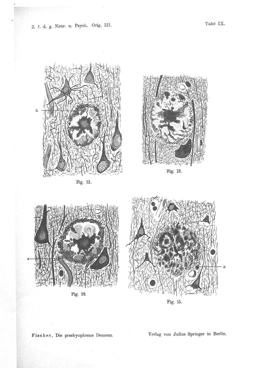
Fischer's seventh stage is the deposit coming apart, and its distinguishing feature is the Scholle — a clod, or an ice floe. He spends a page deciding what the clods are, and the reasoning is careful.
They are not, he concludes, cell remnants, and the argument is quantitative:
"Weiter fällt es auf, daß die Schollen in den Drusen unverhältnismäßig häufiger zu sehen sind als Zellen und Zellreste; wenn nun erstere aus letzteren entstehen würden, müßte man viel mehr Übergänge sehen, als es in Wirklichkeit zutrifft."
("It is further striking that the clods are to be seen in the drusen disproportionately more often than cells and cell remnants; if the former arose from the latter, one would have to see far more transitional forms than in reality one does.")
His alternative is that the clods come from the deposit itself: "daß wenigstens ein Teil der Schollen durch Zerfallsprodukte der Fädchenmassen gebildet wird" — that at least part of the clods are formed by breakdown products of the thread masses. On that reading Fig. 15 is the earlier phase, threads and clods intermixed with clods predominating, and Fig. 13 the later, where only sparse thread remnants remain at the clod margins.
Then, in a single sentence that is easy to read past, he adds a chemical observation:
"Bei der Behandlung nach Marchi werden die in diesen Schollen eingeschlossenen Körnchen bräunlich bis schwarz."
("On treatment by Marchi's method the granules enclosed in these clods become brownish to black.")
Marchi's method — osmium tetroxide with potassium dichromate — is the classical stain for degenerating myelin, and it works because osmium is reduced and blackened by unsaturated lipid that has been chemically altered by degeneration while normal myelin, protected by the dichromate mordant, remains unstained. A Marchi-positive granule is a granule of degenerate lipid.
So Fischer reported, in 1910, that the dissolving plaque contains granular material of degenerate-lipid character, enclosed within larger bodies, arising as the deposit comes apart. He recorded it, drew it, and drew no conclusion from it, because there was none available. His own solvent series two years later would establish that the threads are protein and not lipid — "es kann sich demnach kaum um etwas anderes als eine eiweißartige Substanz handeln" — which makes the lipid granules of Stage VII a discordant finding within his own chemistry, and he does not reconcile them.
Set that against the modern account of what fills the structures at issue. The predominant organelles in the swollen neurites of Alzheimer cortex, on immunogold electron microscopy of human biopsy tissue, are autophagosomes, multivesicular bodies, multilamellar bodies and cathepsin-containing autophagolysosomes (Nixon et al., 2005). A multilamellar body is, structurally, concentric stacked membrane; a multivesicular body is a bounded compartment full of small membrane vesicles; an autolysosome that cannot digest its cargo retains the membranes of everything delivered to it. The lysosome-like organelles that accumulate in swollen axons at plaques are protease-deficient, and therefore predicted to be unable to degrade proteinaceous cargo efficiently (Gowrishankar et al., 2015). And in the aged and diseased brain the phagocytic cell that clears such material becomes a lipid-droplet-accumulating microglion, defective in phagocytosis and proinflammatory (Marschallinger et al., 2020).
The reconciliation is that Fischer's dichotomy had two terms and needed three. He asked whether the clods came from cells or from the threads, and rejected the first because there were not enough intermediate forms between a recognisable cell and a clod. There is a third possibility that produces no such intermediates: the clods are neither a cell nor the fibrillar deposit but the membrane-bounded organelles that were inside a cell, released as units. An organelle spilled from a ruptured neuron does not pass through a series of forms intermediate between a nucleated cell and a granule. It is already a granule, at the moment it arrives.
That reading accounts for four features of the passage at once. It explains why the clods are lipid-positive when the threads are protein. It explains why they outnumber recognisable cell remnants — one lysed neuron supplies a great many organelles and one nucleus. It explains the absence of transitional forms Fischer looked for and could not find. And it explains why he found them at Stage VII, the stage at which the deposit is coming apart and the retraction space fills with granular material: a deposit whose fibrillar component is being removed exposes the non-fibrillar contents that were released with it.
Three cautions are owed. Marchi positivity is a reaction, not an identification, and osmium blackening has other causes. Fischer's clods could contain degenerating myelin from injured axons rather than organelle membrane, and Chapter 15 shows that injured axons are certainly present in the vicinity. And Fischer explicitly hedged — "wenigstens ein Teil", at least a part — leaving open that the clods are heterogeneous, which on this reading they would be. We therefore grade the identification of the clods with released organelle membrane as inference, not as an established correspondence. What is not inference is the observation itself: there is degenerate lipid in the dissolving plaque, it was reported in 1910, and no account of the plaque as a precipitated protein aggregate predicts it.
9. The Nuclei of Unclear Provenance
The second unexplained observation is a sentence about cells, and it is the one on which Part Five turns.
Describing the mature wheel-stage and skein-stage deposits, Fischer writes:
"die kleineren derselben sind immer zellfrei; in den größeren findet man hin und wieder Kerne resp. Kerndetritus, deren Provenienz nicht klar ist… Doch sind solche Vorkommnisse immerhin eine Seltenheit."
("the smaller of them are always cell-free; in the larger ones one finds now and then nuclei or nuclear detritus, whose provenance is not clear… Yet such occurrences are nonetheless a rarity.")
Three claims in one sentence, and they should be separated because they have different strengths.
Smaller deposits are always cell-free. Strong, and consistent with everything else in the scheme: the young deposit lies free in tissue showing no other change.
Larger deposits sometimes contain nuclei or nuclear detritus. A positive observation, in in-situ-fixed human tissue, and therefore reliable in the sense of Chapter 3. He notes that they might be cells enclosed during the deposit's growth, and declines to commit.
Such occurrences are a rarity. A negative frequency claim, and the weakest of the three — though not as weak as his other negatives about cells, because here he is reporting a structure his preparations demonstrably could show, since he found and drew examples of it. On Tafel IX, Fig. 12, the caption records a perishing cell nucleus at the upper left together with an axis cylinder crossing the deposit, and Fischer notes both are "ein sonst seltenes Vorkommnis".
Now set beside it the only modern human measurement of the same thing. In 2001, D'Andrea, Nagele and colleagues examined the localisation of Aβ42 in entorhinal cortex and hippocampus of Alzheimer brains by immunohistochemistry with digital image analysis. They reported that Aβ42 first accumulates in the perikaryon of pyramidal cells as discrete granules that appear to be cathepsin D-positive, suggesting lysosomes or lysosome-derived structures; that regions abundantly populated with such overburdened neurons also contained evidence of neuronal lysis; that lysis resulted in radial dispersion of cytoplasmic contents including Aβ42 and lysosomal enzymes into the surrounding space; and — the sentence that matters here — that a nuclear remnant was found at the dense core of many amyloid plaques (D'Andrea et al., 2001). They supported the reading with two further observations: an inverse relationship between plaque density and pyramidal-cell density, and a correlation between plaque size and the size of local pyramidal cells.
The convergence with the framework of Chapter 6 is close and it predates it by twenty-one years — cathepsin-positive granules in the perikaryon, lysis, dispersal of lysosomal enzymes into the neuropil, one plaque per dead cell. It also, incidentally, supplies the mechanism that the 1990 finding of active lysosomal proteases inside plaques had been waiting for.
But note the collision. Fischer: nuclear remnants inside deposits are a rarity. D'Andrea and Nagele: a nuclear remnant is at the core of many plaques. These are two direct human morphological observations of the same feature of the same lesion, made ninety-one years apart, pointing in opposite directions, on precisely the question that a careful modern evaluation of the intracellular account has to mark as unanswered — the fraction of the human plaque burden formed from within.
Neither figure is usable as reported, and it is not primarily because either observer was careless. Chapter 19 shows that the sampling geometry of a small object inside a larger one, viewed in a thin section, is sufficient on its own to generate a discrepancy of this magnitude and this direction, and that the correction is calculable. It also shows that both observations, corrected, are compatible with a substantial inside-out fraction — and that neither, corrected, establishes one.
Two further asymmetries between the studies should be recorded now because they cut in opposite directions. Fischer surveyed cortex broadly and reports the observation as a general one; D'Andrea and Nagele examined entorhinal cortex and hippocampus, which are exactly the regions where pyramidal amyloid load is highest and the intracellular route should therefore be commonest, and where the largest cell-death events in the disease occur. That biases the modern figure upward relative to the historical one for reasons that are biological and legitimate. Against that, the Nagele line has been little taken up, its proposed uptake mechanism through the α7 nicotinic receptor is a separate and weaker claim that need not be accepted with the morphology, and the 2001 paper reports "many" without a denominator.
10. The Empty Core and the Crowded Rim
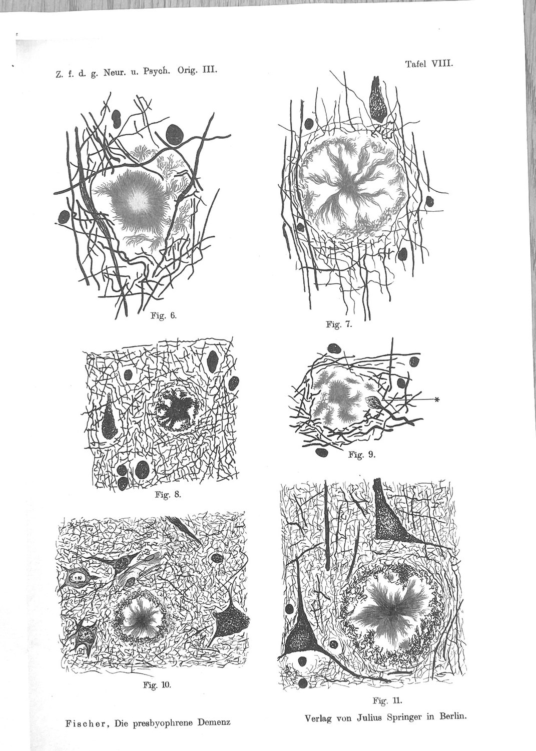
Fischer states the geometry of the mature deposit twice, and in almost the same words each time:
"Die periphere Zone der Drusen enthält demnach häufig ein dichtes Netz von Fibrillen, das Zentrum ist aber meistens frei von Achsencylindern und Nervenfasern, nur ausnahmsweise ziehen auch isolierte Achsencylinder noch durch."
("The peripheral zone of the drusen accordingly often contains a dense net of fibrils, but the centre is mostly free of axis cylinders and nerve fibres; only exceptionally do isolated axis cylinders still pass through.")
A dense fibril net at the rim; a core free of axons; occasional axons crossing, noted as exceptions. This is the dense-core plaque as it is described today — an acellular, afibrillar core with a corona of processes — and Fischer draws it on Tafel VIII.
What he does not do is ask why the centre should be empty. The question is not trivial. If a deposit grew by precipitation from the interstitial fluid into a neuropil that was there beforehand, one would expect the axons to be within it, encased, in the way that a mineral concretion encases the matrix it grew through. Fischer's own vocabulary for the smallest deposits says the axons bend around them — "die Achsencylinder um die größeren Sternchen immer, um die kleinen sehr häufig bogenförmig verlaufen" — but bending around a two-micrometre object is not the same as a fifty-micrometre core with nothing in it.
Two readings produce an empty core, and they are not exclusive.
The first is displacement carried to completion: the deposit grows, the neuropil retracts around it, and the centre ends up empty because everything that was there has been pushed out. This is Fischer's own reading and it is consistent with the Hof.
The second is that the centre was never neuropil. If the core of the deposit is the site where a cell body stood, then the absence of traversing axons is not the result of displacement but of the original occupancy: a pyramidal perikaryon is not a place where axis cylinders run in a dense net, and when it goes, what remains is a space of roughly the right size with the neuropil intact at its margin. This is what the inside-out account predicts, and it predicts something else with it — that the size of the deposit should scale with the size of the cell that produced it. That relation was reported: plaque size correlates with the size of local pyramidal cells (D'Andrea et al., 2001).
The two readings can be told apart, and Fischer's third observation is the one that begins to do it. In the same passage he reports that the halo — the space between core and corona — is "das Gewebe ist ringsherum retrahiert, so daß ein freier Hof herum entsteht", tissue retracted all around so that a free halo arises. Retraction produces a halo outside an object of a given size. It does not produce an empty centre inside a corona of preserved fibrils, because tissue that has retracted is by definition at the margin.
The honest statement is that Fischer's geometry is what an object that arrived at once, at a point, with a diameter set by something other than diffusion, would leave behind — and that it is also compatible with slow displacement. He had no way to distinguish them, and neither does a static image today. What distinguishes them is size distribution, and the prediction is set out in Chapter 31.
11. The Halo Is a Space
The retraction halo is the most misquoted feature of Fischer's description, and the German is unambiguous. Of Fig. 4 on Tafel VII, at the end of Stage II, he writes that the tissue is retracted all round so that a free halo arises. The Hof is what the neuropil vacates. It is not what the deposit adds, and it is not condensed host material.
He was careful about this because he had considered the alternative and rejected it. With Fig. 8 on Tafel VIII, he writes, one can easily gain the impression that the ring bounding the nervous tissue is merely a condensation of the nerve fibrils — "Dagegen sprechen viele Momente." Many considerations speak against it. He gives three, and all three turn on the metachromasia of his own preparation: in Fig. 7 the central core, the strands and the marginal ring present "in gleicher Tinktion und als aus denselben Elementen aufgebaut", in the same tint and as built of the same elements, whereas host fibrils condensed would take the host's colour; in Figs 10 and 11, where the ring is black and the strands brown, the fibrils of the latter pass over into the former or interweave with them, so the two differently coloured parts are one structure; and the threads are much finer than nerve fibrils, with a course "viel wirrer und eckiger", far more tangled and angular. He generalises it negatively at Fig. 16: "Nie aber ist auch nur eine Spur eines Überganges des nervösen Gewebes in die Fädchenmassen zu sehen" — never even a trace of a transition of nervous tissue into the thread masses.
For the present argument the interest is not that Fischer was right about the halo, though he was. It is what the combination of his two findings requires. The material of the ring is the same material as the core, continuous with it, and chemically unlike the fibrillar architecture of the tissue; and the space between the deposit and the intact neuropil is a space the tissue has left. Put together: a body of foreign-looking material occupying a volume from which host tissue has withdrawn, with no continuity anywhere across the boundary.
That is a precise description of an object that was delivered rather than deposited. It is also, on the modern account, a description of the compartment in which the deposit's chemistry does its damage: the microregions of a plaque covered by microglial processes are compact and low in Aβ42 affinity, while uncovered microregions carry high-affinity protofibrillar hotspots, and it is at the hotspots that axonal dystrophy is severe (Condello et al., 2015). Fischer's Hof is the space across which that gradient runs. He could see the geometry and not the chemistry; the modern work supplies the chemistry and inherits the geometry without knowing whose it was.
The unexplained residue is the sharpness of the boundary. A gradient is by definition graded, and a precipitating species falling out of solution should produce a deposit whose edge reflects the concentration profile — diffuse, feathered, and shading into the neuropil. Fischer's Stages III to V do not have that edge. They have a ring. He remarks on it repeatedly and treats it as evidence of the material's foreignness. The modern answer is that the ring is made by a cell: the microglial mantle prevents outward expansion and compacts the deposit, and where the mantle is deficient — in TREM2 R47H carriers, in human autopsy tissue — deposits are less compact and more filamentous and the surrounding neurites suffer, at unchanged plaque number (Yuan et al., 2016). Fischer's sharp edge is a containment boundary. It is one of the few features of his description for which the modern explanation is neither of the two programmes examined here.
12. Glia Around the Older Foci
The fifth of Fischer's defining properties of the process is a negative: the formations "keinerlei reaktive Entzündung hervorrufen", call forth no reactive inflammation whatever. That clause is regularly quoted as a characterisation of the lesion's biology. It is not one. It is the premise of an argument, deployed exactly once, against the hypothesis that the deposit is a filamentous organism, and it appears in a sentence which in the same breath calls the deposit a foreign inclusion and the club-neurites the expression of damage caused by it.
What is much less often noticed is that Fischer, two years later, reported a glial reaction — and reported it stage-dependently. In the 1912 paper, having again rejected a glial origin for the deposits, he records:
"außerdem fanden sich besonders um die größeren und älteren Herde gewucherte und vergrößerte Gliazellen."
("in addition there were found, especially around the larger and older foci, proliferated and enlarged glial cells.")
The qualifier is the finding. Not "glial cells were present around the deposits" but around the larger and older ones — the same conditionality he had established for the neurites in 1910, applied to a different cell, and stated as a positive observation rather than as an absence.
Three things follow.
Fischer's "no inflammation" and Fischer's gliosis are not in conflict, and the distinction is the one he intended. Entzündung in 1910 meant an exudative, leucocytic reaction of the kind a fungal colony would provoke — the thing whose absence refutes an infection. Reactive hypertrophy and proliferation of glia is not that, and he reports it without embarrassment. Reading the anti-fungal clause as a claim that the plaque provokes no cellular response inverts its function in the text.
The stage-conditionality is right, and it was measured again a century later. Quantitative neuropathology in temporal neocortex across forty subjects with symptom durations of four to twenty years found that plaque burden is essentially stationary while per-plaque features change: dystrophic neurites labelled with SMI312 rose with symptom duration (Kendall τ = 0.34), GFAP-positive reactive astrocytes rose (τ = 0.30), and CD68-positive microglial activation rose steeply (τ = 0.48), while IBA1-positive microglial number did not move at all (τ = 0.045) (Serrano-Pozo et al., 2016). Fischer's proliferated and enlarged glial cells around the larger and older foci is the GFAP arm of that result, reported in 1912 on the correct conditional.
And what he could not see is exactly the cell the modern account needs. Bielschowsky silver does not demonstrate microglia. The astroglial reaction was available to him through the neuroglia stains in his panel; the microglial mantle was not. This matters for the argument of Part Five, because the cell that arrives after the rupture in the PANTHOS sequence — glia invade — is the one cell class in the whole structure that Fischer's preparations could not show. His statement that nuclei inside deposits are rare is therefore a statement about a population from which the most numerous plaque-associated cell type has been silently removed. Chapter 19 uses that.
13. The Colour Clock and the Direction of Growth
Running underneath the stage table is an instrument, and it is the most original thing in the 1910 monograph.
"Wir sehen nämlich, daß die Sternform als die jüngste Bildung immer nur schwarz gefärbt erscheint, wogegen die Morgensterne und die Strähnchen sehr häufig eine bräunliche Färbung annehmen; wir könnten also daraus auch schließen, daß sich die Fädchen in den jüngsten Stadien schwarz, in den späteren braun färben."
Threads impregnate black when young and brown when older. Fischer then applies the rule within a single deposit:
"Das gleiche Verhalten bemerkt man nun auch im Stadium der Rädchenbildung; die zentralen Partien sind daselbst meist bräunlich oder rötlich tingiert, wogegen der Randring immer schwarz erscheint… daraus folgt, daß auch auf Grund des tinktoriellen Verhaltens der Randring als jüngste Bildung der Drusen erscheint."
In a wheel-stage druse the centre is brown and the rim is black. If black is young, the rim is the most recently formed part, and therefore the deposit grows outward from the centre, adding new material at its margin. That is a growth model derived from a staining property, cross-checked against an independent observable — the size series — and consistent with it. It agrees with what in vivo two-photon imaging now shows directly: plaques appear and then enlarge outward from an established core.
The relevance here is the corollary Fischer did not draw. If the core is the oldest part of the deposit and material accretes at the rim, then whatever the deposit began as is still at its centre, and the centre is the place to look for the deposit's provenance. Every feature of the modern intracellular account concentrates its evidence at the same address: the nuclear remnant is reported at the dense core (D'Andrea et al., 2001); the perinuclear accretion of fibrils around a disintegrating nucleus is the penultimate step of the PANTHOS sequence (Lee et al., 2022); and the lysosomal proteases that were the framework's founding anomaly are contents of the cell, which on rupture would be delivered at the point of rupture and then buried by subsequent accretion.
This gives the colour clock an application it has never had. Fischer's inference makes the core a stratigraphic object: the oldest material in a deposit, laid down first, and progressively sealed in by later growth. If plaques form by two routes, the routes should differ most at the core and least at the rim, because the rim is where both routes look the same — accreted fibril from the interstitium. A provenance signature, if it exists, is a core signature, and it is being progressively covered by a process Fischer described.
The limitation he does not state should be stated. Silver impregnation intensity depends on section depth, fixation, local pH and packing density as well as on the age of the substrate, and a colour difference between core and rim could reflect packing rather than chronology. His defence is indirect and real: the same colour ordering holds across deposits of different sizes as it does within one, and two independent orderings agreeing is better than either alone. It is not a demonstration, and we grade the growth-by-accretion claim as inference throughout.
Part Four — The Inverted U
14. The Rule, Whole
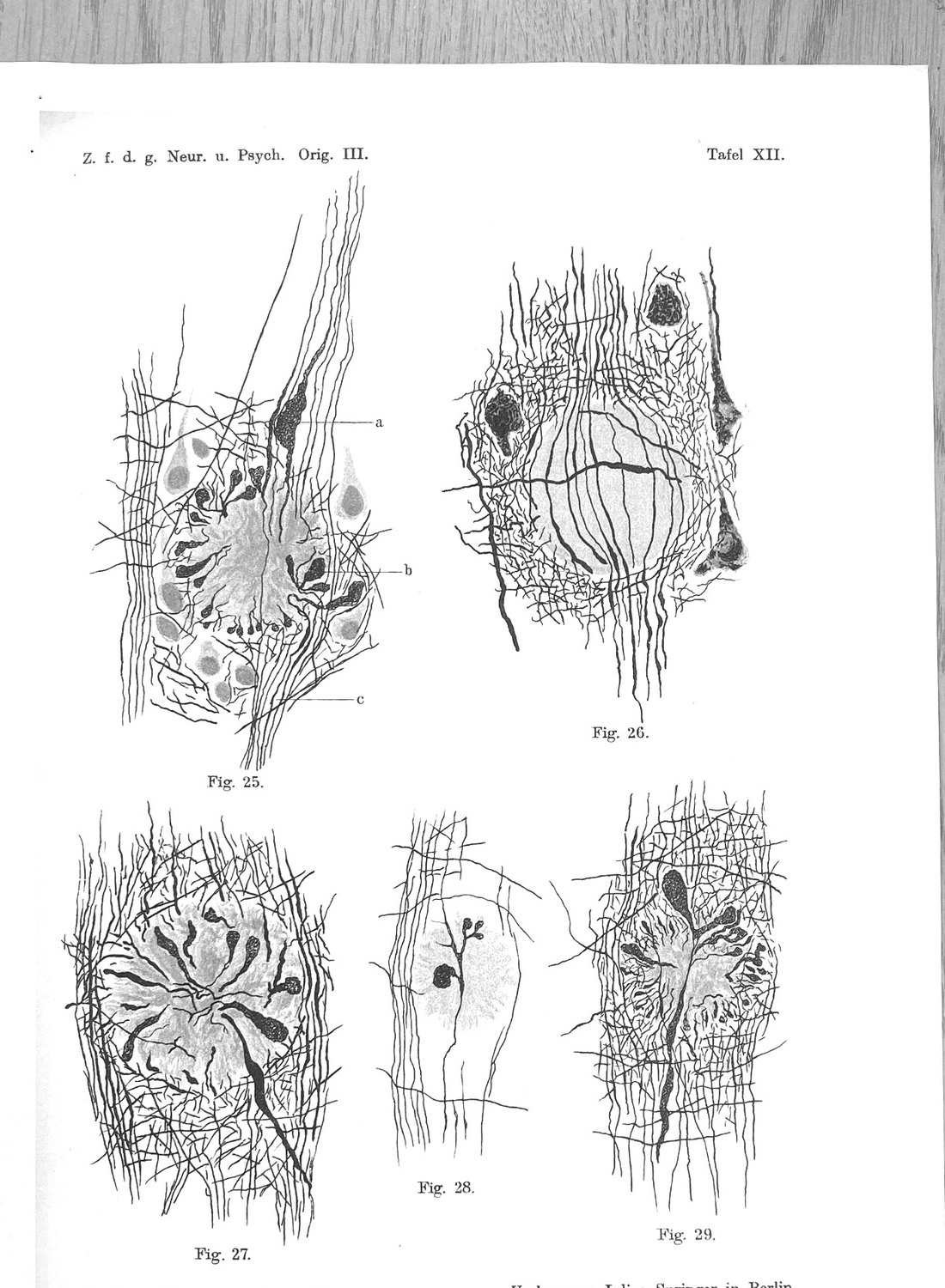
The most consequential sentence Fischer wrote is on page 381 of the 1910 monograph, and it is rarely quoted to the end.
"Die kolbigen Wucherungen der Achsencylinder kommen nur in etwa 50% der Fälle vor; dabei auch nur um die größeren Drusen; am häufigsten finden sie sich um das Stadium V, dann um Stadium IV, seltener um Stadium III, nie in der Nähe der Stadien I und II, ebenso auch nie um Stadium VIII. (Siehe Einteilung der Stadien auf Seite 392.) Warum sich in den einzelnen Fällen die Keulen vorfinden, in den anderen nicht, ist nicht klar geworden, im histologischen Verhalten war sonst keine Differenz merkbar."
("The club-shaped proliferations of the axis cylinders occur in only about 50% of cases; and then only around the larger drusen; most frequently they are found around Stage V, then Stage IV, more rarely around Stage III, never in the vicinity of Stages I and II, and likewise never around Stage VIII. (See the classification of stages on page 392.) Why the clubs are present in individual cases and not in others has not become clear; in histological behaviour no other difference was perceptible.")
Four features of that sentence bear on the argument.
It is a per-lesion measurement with the brain as its own control. Every deposit in a section is an independent observation scored for two variables — its stage, and whether clubs surround it — in the same tissue, the same subject, the same fixation, the same stain. Age, agonal state, post-mortem interval, disease duration and genotype are held constant by construction. This is the design quantitative neuropathology now regards as strongest for questions of local pathology, and Fischer used it because it was the only way to extract an ordering from static material.
The relation is an inverted U. Clubs peak at Stage V, decline through IV to III, are absent at I and II — and are absent again at VIII. The second half of that is not in the secondary literature. It changes the shape of the claim from a ramp to a curve, and the curve is what makes it informative.
The percentage attaches to cases, not certainly to plaques. "der Fälle" in Fischer's German most naturally means cases, and the per-deposit restriction is carried by the clause that follows, "dabei auch nur um die größeren Drusen." The German will bear either reading and we do not resolve it, because the argument does not need it: the stage-ordering is unambiguous either way. What can be said without ambiguity is that in half his cases the deposit provoked no visible neuritic reaction anywhere, and that where it did, it did so around the older and larger forms.
And Fischer marks the residual as unexplained. He has a variable that explains part of the variance, says so, and then says the rest is unaccounted for. That is what separates his staging from a taxonomy: he expected the scheme to predict something, checked whether it did, and recorded the shortfall.
He also states the rule as a defining property of the process rather than as an incidental finding. In the five-part definition on pages 391–392, the formations are those which lead to proliferations of the axis cylinders "in einem kleinen Prozentsatz der Fälle und in bestimmtem Alter des Prozesses" — in a small percentage of cases and at a definite age of the process. Stage-conditionality of injury is written into his definition of what the lesion is.
And there is a regional replication, which the mistranslation of his distribution sentence threw away. Fischer found drusen in the cerebellum in two of ten cases whose cortex was richly beset, and described them: "Im Kleinhirn waren die Drusen immer nur sehr spärlich vertreten, saßen nur in der Mitte der Molekularschichte, zeigten meist das Stadium III… und enthielten nie Keulen." Sparse, confined to the middle of the molecular layer, mostly Stage III, and never containing clubs. Where the deposits stay young, no clubs form — in a region as well as in a field. That is the stage rule tested on a second axis, and it holds.
15. What Is Inside a Club
Fischer defined the injurious form of the lesion by a structure whose contents he could not examine. Three modern answers exist for what is inside it, they were obtained by different methods in different material, and they have never been arbitrated.
Answer one: amyloid fibrils in synaptic compartments. Three-dimensional reconstruction using a β-sheet-binding dye showed fibrillar amyloid inside individual synaptic compartments, associated with abnormal morphology and in places appearing to pierce the membrane; in dendrites, rising intraneuronal fibrillar signal tracked falling neurofilament (Capetillo-Zarate et al., 2011). Intraneuronal Aβ42 in human tissue localises to multivesicular bodies within synaptic terminals, associated with abnormal synaptic structure before plaque pathology (Takahashi et al., 2002), and its accumulation is associated with early changes in MAP2 in neurites and synapses (Takahashi et al., 2013). On this account the club is a compartment distended by cargo it retained.
Answer two: stalled autophagic vacuoles. Immunogold electron microscopy with compartmental markers on human cortical biopsy tissue found autophagosomes, multivesicular bodies, multilamellar bodies and cathepsin-containing autophagolysosomes to be the predominant organelles, in very large numbers; on quantification, autophagic vacuoles comprise over ninety-five per cent of the organelle content of grossly swollen axonal segments (Nixon et al., 2005). On this account the club is a segment of axon in which compartments generated normally and transported retrogradely toward the perikaryal lysosomes have stalled. The causal claim is supported by the observation that inhibiting endolysosomal acidification produces neuritic dystrophy in wild-type mice with no amyloid deposition at all.
Answer three: protease-deficient lysosomes, stalled by the deposit. An independent laboratory documented a massive accumulation of lysosome-like organelles at plaques, the majority within swollen axons contacting the deposits, present from the earliest stages of deposition; the organelles lack multiple soluble luminal proteases, and BACE1 accumulates at the same sites (Gowrishankar et al., 2015). The authors' interpretation is that the extracellular deposit causes a local impairment of retrograde transport of lysosome precursors.
The three are much closer in what than in why. Multivesicular bodies appear on all three lists; a compartment full of retained cargo and a compartment full of uncompleted autophagic vacuoles are not cleanly separable in a neurite that is doing both. The disagreement is about direction. Answers one and two make the club a manifestation of a cell-autonomous failure that the deposit accompanies; answer three makes it a local injury the deposit inflicts.
Fischer's rule bears on this, and it is the reason the inverted U is worth recovering.
A deposit that injures a passing neurite by chemical action at its edge should produce a monotone relation: more deposit, more mature deposit, larger surface, longer residence, more injury — rising to a plateau at most. That is what answer three predicts, and it is what the field has assumed for a century. The observed relation, on the only measurement ever made of it, is not monotone. It rises from zero at Stages I and II, peaks at V, and returns to zero at VIII.
A curve with two zeroes requires two different explanations for its two ends, and both of Fischer's are explanations about the neurite, not about the deposit.
At the young end the deposit is present and the reaction is absent. Fischer's deposits at Stages I and II are two to thirty micrometres, lying free, in tissue showing no other change. Something must be crossed before the neurite responds, and on the modern accounts the threshold is a cargo threshold: the compartment must be loaded enough to distend.
At the infiltrative end the reaction is absent because the neurite is gone. Fischer is explicit: within the infiltrated areas of Stage VIII only the thicker traversing axis cylinders take the stain at all, "wogegen die größte Mehrzahl derselben ungefärbt, aber bei stärkerer Abblendung noch deutlich sichtbar ist" — the great majority unstained, though still visible under strong stopping-down. And in the same passage: over areas sometimes larger than an immersion field, the axis cylinders and the fibrillar network have vanished completely, and of the nervous tissue only the ganglion cells persist, severely shrunken and having lost most of their processes.
There is no club because there is no longer an axon in a condition to make one. That single clause is the strongest morphological statement in the entire Fischer corpus about the nature of the club, and it says the club is not damage inflicted on a passive structure. It is something an intact, transporting, cargo-handling process does, and it requires the process to be alive enough to do it. A dead axon does not swell. A jammed one does.
That is a statement both intracellular programmes need and neither has made from human material. It is also in direct tension with the third answer above, since a deposit that stalls transport by chemical injury from outside should stall it most where the deposit is most confluent — which is Stage VIII, where the clubs are absent.
One caveat must be entered against reading the inverted U as established, and it is a real one. Gowrishankar and colleagues found lysosome accumulation in axons at plaques from the earliest stages of deposition. If that transfers to human tissue, then Fischer's young-end zero is a limit of his stain rather than a biological threshold: the organelle accumulation is there at Stages I and II and the axon is not yet swollen enough for silver to show a Keule. Both readings — a genuine threshold, or a detection floor — predict the same appearance in Bielschowsky and are distinguished only by a marker Fischer did not have. Chapter 17 sets out the test.
16. Why the Two Ends Fail Differently
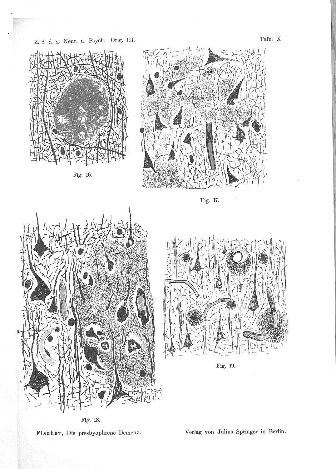
The eighth stage is the most thoroughly misread item in Fischer's scheme, and its correct reading is what makes the inverted U mechanistically interesting rather than merely curious.
Immediately after printing the stage table Fischer writes:
"Die Stadien I–V sind so aufzufassen, daß I das jüngste, V das älteste darstellt. Die drei letztgenannten Stadien lassen sich schwieriger abschätzen, doch scheint mir Stadium VI und VII zu den ältesten Drusen zu gehören; das Stadium VIII möchte ich wieder als einen jüngeren Prozeß ansehen, gegen dessen Fortschreiten das Gewebe weniger Widerstand aufbringen konnte, so daß ein diffuses Infiltrieren zustande kam."
Stage VIII is a younger process, against whose advance the tissue could muster less resistance. Not the terminus; the second-youngest thing in a list ordered by age. He supports it with the colour clock — the infiltrative masses stain only black, the young colour — and hedges the inference in his own emphasis: "was auch dafür sprechen könnte (ich betone hier das „könnte")".
And he gives the mechanism in one clause: the tissue does not retract because it is not given time to. "das infiltrative Durchwuchern, als Ausdruck einer sehr schnellen Wucherung, bei der das nervöse Gewebe keine Zeit zur Retraktion mehr hatte" — infiltrative overgrowth, as the expression of a very rapid proliferation in which the nervous tissue no longer had time to retract.
So Fischer's eighth stage is not a measure of how long the disease has run. It is a statement about the ratio between the rate of deposition and the capacity of the host to accommodate it — the nearest thing in his framework to a variable of resistance, and the one place in the scheme where the tissue is allowed to have a say.
The two ends of the inverted U are therefore failures of opposite kinds, and this is the point at which Fischer's morphology and the modern cell biology say the same thing in different vocabularies.
Table 2 — The two zeroes of the inverted U
| Stages I–II | Stage VIII | |
|---|---|---|
| Fischer's description | Smallest forms, 2–30 µm, lying free in tissue "nicht wesentlich verändert" | Confluent masses replacing the neuropil over areas larger than an immersion field |
| State of the axon | Intact; bends around the deposit | "die größte Mehrzahl… ungefärbt" — the great majority gone |
| Why no club | The stimulus has not reached threshold | There is no process left to swell |
| Fischer's own term | Youngest stage of the growth series | A younger process in tissue offering less resistance |
| Rate relative to accommodation | Slow enough for retraction; halo forms | Too fast for retraction; no halo |
| Modern reading | Cargo load below the distension threshold | Deposition outruns containment; the deposit is uncompacted |
The right-hand column is where the modern correspondence is sharpest and least expected. Because Fischer's series runs from small and simple to large and complex, and because injury rises along it, the natural reading is that compaction is deterioration. It is not. Compaction is what the tissue achieves: microglial processes form a barrier that compacts the deposit and holds high-affinity protofibrillar species off the neuropil, and where coverage is deficient the deposit is filamentous and the neurites are destroyed (Condello et al., 2015; Yuan et al., 2016). Fischer's Stages III to V describe a deposit being brought under control, and the clubs alongside them are the cost of a containment that is under way and incomplete. His Stage VIII — fast, uncontained, in tissue that resists less — is the failure mode, and it carries no clubs because it does not leave enough axon to swell.
The clinical correlate Fischer attached to the infiltrative mode fits the same reading. In 1912 he sets Case 12, an acute delirium of a few days' onset with abundant smallest star-drusen and massive infiltrates and no older forms at all, beside an earlier case, and draws the inference in spaced type: an acute fresh sowing produces a delirium that can remit; the drusen persist and mature; and a further Schub — a wave — of thread formation produces a new delirious state. Whatever one makes of a two-case model, it is a model in which the rate of arrival is a clinical variable independent of the total accumulated, which is what Stage VIII is.
There is a modern analogue of the rate variable in Gouras's programme and it has the right sign, though it belongs to a different level of the system. Chronic synaptic inhibition, applied by two independent means, reduced plaque burden and worsened synaptic and memory outcomes in model animals, with the deterioration occurring in the setting of reduced deposits and elevated intraneuronal amyloid (Tampellini et al., 2010). Deposit burden and functional outcome were driven in opposite directions in the same animals. That is a designed demonstration that the tally of deposits is not the variable that tracks function — which is the same conclusion Fischer reached from the other end, by finding that half his cases had no neuritic reaction at all.
17. The Test That Would Settle It
The inverted U is the sharpest untested claim in this literature, and it is testable in existing human tissue with existing reagents. Setting out the design precisely is worth more than another argument.
The unit is the individual deposit, not the field and not the case. Every deposit in a section is scored, and the brain serves as its own control, which is what removes age, genotype, agonal state, post-mortem interval and disease duration from the comparison.
Four terms are measured on each deposit. Compaction: the ratio of dense-core area to total deposit area, by thioflavin S or an equivalent conformation-selective stain. Mantle coverage: the fraction of the deposit perimeter, in a confocal optical section through its widest plane, in contact with microglial process membrane; IBA1 for the process label, with a P2RY12 counter-stain, since plaque-associated cells characteristically lose the homeostatic marker. The denominator must be deposit perimeter, not field area, or the measure degenerates into a microgliosis score. Halo extent: the radial distance from the compact core at which conformation-selective staining for protofibrillar species falls to background. Neuritic dystrophy: the area of SMI312- and LAMP1-positive dystrophic profiles within a fixed radius of the deposit margin, normalised to perimeter.
The prediction is a curve, and its shape is the result. Dystrophy should rise from near zero at the uncompacted, mantle-free extreme, peak at intermediate compaction with incomplete mantle, and fall again at the diffusely infiltrative extreme where the neuropil has been replaced. The claim is falsified if the relation is monotone. A monotone relation is what a corrosive-deposit model predicts and would leave the modern intracellular accounts without their oldest morphological support.
Two markers must be carried, because they separate the two readings of the young-end zero. Fischer could score only silver-visible axonal swelling. A modern series should score, on the same deposits, both a coarse morphological dystrophy marker and a lysosomal marker — LAMP1, or cathepsin D. If LAMP1-positive profiles are present around Stage-I-equivalent deposits where SMI312-positive swellings are not, the young-end zero is a detection floor and the organelle response precedes the morphology, as Gowrishankar and colleagues report from mouse. If both are absent together, the threshold is real. This single comparison decides between two readings that have been conflated since 1910, and it costs one extra channel.
The confounds are known and each biases in a stated direction. Agonal state and terminal illness alter microglial morphology. Post-mortem interval degrades fine process detail before somata, biasing mantle coverage downward — the direction that flattens a true effect, so a null obtained without matching it should not be read as a null. Fixation duration alters conformation-selective epitope retrieval, bearing on the halo term. Fischer controlled the second of these better than most modern series, by fixing in situ within minutes of death, and a modern series should say what its interval was.
And one term should be reported that no scheme currently reports: the stage histogram. Fischer observed that no brain contained all stages and none contained only one, and that regions could differ in stage composition. If deposition proceeds in waves, the distribution of deposit states within a brain is informative independently of the mean: a tightly clustered distribution indicates one wave, a bimodal one indicates two. The mean is what a staging scheme reports and the histogram is what the wave model calls for. It has never been taken.
The value of this experiment does not depend on which programme is right. If the curve is inverted-U, the club is a reaction of a living process with a window, and accounts in which the deposit corrodes its neighbours lose their principal structure. If the curve is monotone, the oldest quantitative claim in this field is wrong and should be retired, which is also worth knowing. What is not defensible is the present position, in which a hundred-and-sixteen-year-old measurement that discriminates between two classes of mechanism has never been repeated.
Part Five — The Fraction
18. A Rarity and a Many
The question the intracellular accounts cannot answer from their own material is what share of the human plaque burden formed from within. It is not a small question. If a substantial fraction of plaques are the residue of dead neurons, then plaque burden is partly a record of past cell death, and its long-standing failure to correlate with cognition is what one would expect of a variable that is part cause and part gravestone. If the fraction is negligible, the intracellular route is a mouse phenomenon.
The obstacle is stated candidly by the programme itself: a finished deposit is the same object whether it condensed from secreted peptide in the neuropil or was released by a ruptured cell, the peptide carries no provenance label, the route by which a plaque formed is erased by its completion, and the intracellular route destroys the very cell whose identity would be needed to attribute it.
There is, however, one thing a cell has that a precipitate does not, and which is not made of the peptide. It has a nucleus. And the nucleus is the only component of the neuron that is both large enough to be seen in a section, distinctive enough not to be confused with the deposit, and — critically — not consumed by the process that produces the deposit. In the PANTHOS sequence the fibrils accrete in the perinuclear region around a nucleus that is visibly disintegrating but still present in the great majority of these cells (Lee et al., 2022). A nuclear remnant at the centre of a plaque is the closest thing to a provenance signature the lesion offers.
Two human observations of that signature exist. They were made ninety-one years apart and they point in opposite directions.
Fischer, 1910, on the mature deposits: the smaller ones "sind immer zellfrei", are always cell-free; in the larger ones one finds "hin und wieder Kerne resp. Kerndetritus, deren Provenienz nicht klar ist", now and then nuclei or nuclear detritus of unclear provenance; and "Doch sind solche Vorkommnisse immerhin eine Seltenheit" — yet such occurrences are nonetheless a rarity.
D'Andrea, Nagele and colleagues, 2001, on entorhinal cortex and hippocampus: "A nuclear remnant was found at the dense core of many amyloid plaques, strengthening the idea that each amyloid plaque represents the end product of a single neuronal cell lysis."
Neither figure has a denominator. Neither study was designed to measure this. And on the face of it they contradict each other on the one observable that bears on the open question.
We argue in the next chapter that they do not contradict each other, that both are uncorrected two-dimensional profile counts of a three-dimensional coincidence, that the correction is arithmetic, and that after it both are compatible with a wide range of values for the fraction and neither establishes one. What we do not argue is that Fischer's observation is negligible. It is the oldest measurement of the inside-out fraction in existence, it was made on in-situ-fixed human tissue with a minimal post-mortem interval, and it has never been cited in this connection by anybody.
19. The Geometry of the Remnant
A nuclear remnant is a small sphere inside a larger sphere. A histological section is a slab. The question "how often does a plaque profile contain a nuclear profile" is therefore not a biological question in the first instance; it is a question about the probability that a randomly positioned slab which intersects the outer sphere also intersects the inner one.
Let the deposit be a sphere of diameter D and the nuclear remnant a concentric sphere of diameter d, and let the section have thickness t. A slab of thickness t intersects a sphere of diameter D for a range of positions of length D + t, and intersects the inner sphere for a range of length d + t. Among sections that show the deposit at all, the fraction that also show the remnant is therefore
P = (d + t) / (D + t)
This is the whole correction, and it is worth noticing how strong it is. Even if every plaque in the tissue contained a nuclear remnant, only a minority of plaque profiles would show one.
Table 3 — Fraction of plaque profiles containing a nuclear profile, given that every plaque contains one
| Section thickness t | Deposit D = 30 µm | D = 50 µm | D = 70 µm |
|---|---|---|---|
| 6 µm (modern paraffin) | 0.39 | 0.25 | 0.18 |
| 15 µm (thick silver section) | 0.51 | 0.35 | 0.27 |
| 40 µm (free-floating / confocal) | 0.69 | 0.53 | 0.44 |
Values computed with d = 8 µm, a condensed pyramidal nuclear remnant. Section thickness for Fischer's preparations is not stated in the passages examined; silver impregnation of the period was typically performed on sections of the order of ten to twenty micrometres, and the middle row is the appropriate comparison.
Three consequences follow immediately.
The ceiling on any two-dimensional profile count is low. Under realistic parameters a 2-D count underestimates the true fraction by a factor of two to five. A study reporting that thirty per cent of plaque profiles contain a nuclear remnant is compatible with every plaque containing one. A study reporting that few do is compatible with a majority containing one.
Geometry alone spans the distance between the two reports. The ratio between the most and least favourable cells of Table 3 is about four. Fischer scored mature deposits across cortex generally; D'Andrea and colleagues scored entorhinal and hippocampal plaques, in regions where pyramidal amyloid load is highest and where the largest cell-death events in the disease occur, using immunohistochemistry with nuclear visualisation and digital image analysis. Before any difference in detection efficiency is considered, the two studies are not sampling the same quantity.
And detection efficiency multiplies the geometric term, in a direction that is not symmetric between the two studies. The observed rate is f × P × ε, where f is the true fraction of deposits formed from a cell and ε is the probability that a remnant intersected by the section is recognised as one. Bielschowsky is a fibrillar silver impregnation; nuclei appear through counterstain, the remnant is by hypothesis disintegrating, and Fischer states plainly that when he saw one he could not determine its provenance. Digital image analysis of an immunostained section with a nuclear counterstain is a substantially more sensitive instrument for this specific object. Put f = 0.5, P = 0.30, and ε = 0.3 for the historical preparation, and the expected observed rate is about five per cent — which is what a careful observer would call eine Seltenheit. Put the same f, the same P, and ε = 0.8 for the modern one, and it is about fifteen per cent — which an author might reasonably call many. The same underlying fraction generates both descriptions.
Two further corrections belong here, and they run in opposite directions.
Against the intracellular reading: the nucleus is not the only nucleus in the neighbourhood. A dense-core plaque carries a mantle of microglia, and astrocytic processes converge on it. Any nucleus seen inside a plaque profile may be a visiting cell rather than a resident corpse — and this is precisely the ambiguity Fischer recorded when he wrote that the provenance was not clear. His preparations could not have resolved it, because his stain did not demonstrate microglia and his counterstain did not distinguish cell classes. It follows that his figure is an upper bound on the observed rate of neuronal remnants — some of what he counted was not neuronal — which lowers the numerator in the estimate of Chapter 20 without changing the direction of the bound that estimate places on f.
For it: the discriminator is position, and it is available. The colour clock of Chapter 13 makes the core the oldest part of the deposit and the rim the youngest. A nucleus at the core was there before the material around it was laid down; a nucleus at the mantle arrived afterwards. This is why the 2001 report specifies "at the dense core" and why that specification is the load-bearing word in the sentence. Any modern count must score radial position, and a count that does not is uninterpretable.
Finally, an observation that at first appears to contradict the whole reading and on inspection sharpens it. Fischer says the smaller deposits are always cell-free and only the larger ones ever contain nuclei. Geometry predicts the opposite: P rises as D falls, so small deposits should show remnants more often, not less. The resolution is a matter of scale. Fischer's smallest forms are "kleinste Krystallsternchen" of about two micrometres; Stage II runs from eight to thirty. A pyramidal perikaryon is fifteen to twenty-five micrometres across and its contents are correspondingly bulky. A two-micrometre deposit cannot be the residue of a lysed neuron; there is not enough of it. His small deposits are therefore not candidates for the intracellular route on size grounds alone, and their being uniformly cell-free is exactly what should be observed.
That yields the structural claim of this Part, which is stronger than the numerical one and independent of it. If both routes are real, Fischer's stage series is not one developmental sequence but two overlapping populations conflated by a morphological ordering. A seeded extracellular deposit begins at two micrometres and grows by accretion — the process the colour clock describes. A cell-derived deposit does not begin at two micrometres; it arrives at once, at roughly cell scale, and enters the series at the size of Stage IV or V without ever having passed through I or II. The two populations would be indistinguishable in a silver preparation, would be ordered by size into a single apparent sequence, and would differ systematically only in what sits at their centres.
20. What Fischer's Numbers Bound
It is worth being exact about what the historical observation can and cannot be made to yield, because the temptation in both directions is real.
It cannot yield the fraction. f is not identifiable from a single uncorrected profile count with unknown t, unknown ε and an unknown mixture of resident and visiting nuclei. Anyone who converts "eine Seltenheit" into a percentage is inventing three parameters.
It bounds the fraction from below, not from above, and that is the surprise. Write the observed rate as f × P × ε. If Fischer's rarity is read as roughly five per cent of mature deposit profiles, and P ≈ 0.35 for his sections, then f × ε ≈ 0.14; and since ε ≤ 1, it follows that f ≥ 0.14. That floor is reached only if his detection were perfect, which it plainly was not. At ε = 0.3 the estimate is f ≈ 0.5; at ε = 0.14 it reaches unity, at which point the data cannot exclude a universal intracellular origin for mature deposits. Read as a rate rather than as an adjective, Fischer's Seltenheit is evidence for a substantial intracellular fraction rather than against one — which is the reverse of how a negative-sounding observation naturally reads, and the reason it has been worth recovering.
Two things weaken that inference and both should be stated. Reading "eine Seltenheit" as five per cent is an assumption, and nothing in the text fixes the number; at one per cent the floor falls to about three per cent. And some share of the nuclei he saw will have been microglial or astrocytic rather than neuronal remnants, which lowers the numerator — an effect that cannot be estimated from his material at all, because his stain did not show him the cells that would have to be subtracted. The floor is therefore soft. It is not, however, a ceiling, and it has occasionally been read as one.
It does constrain the small-deposit population strongly. "Die kleineren derselben sind immer zellfrei" is a statement with P at its most favourable — a small deposit is the easiest case in which to catch a central nucleus — and Fischer reports a categorical absence. This is his most informative datum on the question, and it says that the population of small fibrillar deposits is not made of dead cells. That is a real constraint and it is consistent with what both modern programmes would predict.
And it says nothing whatever about the diffuse deposit, which his stain did not show. Chapter 23 takes that up.
Two general lessons deserve stating, because they apply beyond this instance.
The first is that a morphological negative from a historical preparation is a composite of biology, geometry and reagent, and the three are separable only when the section thickness, the object dimensions and the staining behaviour are specified. None of the three is specified in the classical literature, which is why a century of citation of Fischer's negatives — on regional distribution, on inflammation, on cells in plaques — has repeatedly converted a conditional observation into an absolute one.
The second is that the modern literature has the same problem and has not noticed. The 2001 report of nuclear remnants at the core of many plaques is a profile count with no denominator, no stated section thickness and no radial-position analysis, and it has been cited for twenty-five years as either strong support for neuronal lysis or as an unreplicated curiosity, depending on the citing author's commitments. It is neither. It is an uncorrected measurement of a quantity that is straightforward to measure correctly and that nobody has measured correctly.
21. A Countable Design
The question is answerable, in human tissue, with existing reagents, in one study. The design follows directly from Chapter 19 and is set out here in enough detail to be criticised.
Abolish the geometric term by working in three dimensions. Score whole deposits, not deposit profiles. Either serial-section reconstruction or thick-section confocal imaging through the full depth of each deposit removes P from the estimate entirely: a deposit either contains a nuclear remnant or it does not, and the observed rate becomes f × ε directly. Section thickness must exceed the deposit diameter, which for dense-core plaques means optical sectioning through at least eighty micrometres. This single change recovers a factor of two to five that no amount of additional two-dimensional counting can recover.
Score radial position, and treat it as the primary discriminator. Each nucleus found is assigned a normalised radial coordinate from the deposit centroid to its margin. The prediction of the intracellular route is a core-weighted distribution; the prediction for visiting glia is a mantle-weighted one. A mixture is expected and is informative: the core-weighted component estimates f, and the mantle-weighted component provides an internal control for detection efficiency, since plaque-associated microglia are numerous and their nuclei are unambiguously present.
Discriminate cell class, and accept that the neuronal marker will fail. IBA1 or P2RY12 identifies microglia and PU.1 their nuclei; GFAP or ALDH1L1 identifies astrocytes. The neuronal identification is the hard part, because a disintegrating nucleus loses NeuN and the surrounding cytoplasm is by hypothesis gone. The workable definition is therefore negative and positional: a chromatin-containing nuclear profile at the deposit core that is not attributable to any identifiable glial class. This is a weaker criterion than one would like and it should be stated as such; it errs toward over-counting, and the mantle-weighted glial component bounds the error.
Stratify by deposit size, and test the mixture prediction. If the stage series is two conflated populations, the frequency of core-weighted nuclear remnants should be near zero in small deposits, rise sharply at a threshold corresponding to roughly one cell's volume of material, and be approximately flat above it. If instead deposits form by a single accretive process, the frequency should be a smooth function of size with no threshold. The mixture claim is falsified if the size–frequency relation is smooth. This is the sharpest test in the paper and it comes out of Fischer's observation that small deposits are always cell-free.
Stratify by region, and use the cerebellum as the negative control. Fischer found cerebellar deposits sparse, confined to the middle of the molecular layer, mostly Stage III and never carrying clubs. That layer is where Purkinje dendritic arbours lie, and a 2002 human study independently proposed that cerebellar diffuse plaques represent the remnants of destroyed Aβ42-laden segments of Purkinje dendritic trees, especially at their bifurcations (Wang et al., 2002). The two accounts of the same deposits are ninety-two years apart and agree on the layer. Whether they agree on the mechanism is testable: dendritic remnants would carry no nucleus, so cerebellar deposits should be core-nucleus-negative even if the intracellular route is general. A design that finds nuclear remnants in cerebellar molecular-layer deposits has found something wrong with its own criterion.
Report what a null would mean. If core-weighted nuclear remnants are found in a negligible fraction of whole mature neocortical deposits, with the glial internal control confirming adequate detection, then the intracellular route contributes little to the human plaque burden, and the strongest version of the inside-out claim should be withdrawn. That outcome is a real possibility and the design should be powered to declare it.
We note, without attributing motive, that this experiment requires no new reagent, no animal, no cohort and no instrument that a well-equipped neuropathology department has lacked for twenty years. What it requires is that somebody regard the question as answerable. The most careful published evaluation of the intracellular framework grades the fraction of human plaque burden formed inside-out as beyond the evidence and lists fate-mapping in knock-in mice as the route to settling it. Fate-mapping settles it in mice. The nuclear remnant settles it, imperfectly but directly, in people — and the first measurement of it was made in 1910.
Part Six — Where the Legacy Does Not Hold
22. Stage VI Belongs to the Other Route
An argument that finds only convergence is not an argument. Fischer's sixth stage is the place where the intracellular accounts have least to say and the extracellular route is strongest, and the asymmetry is instructive.
Fischer's own word for the vascular deposit is Pelzbesatz, fur trim, and it appears in his case protocols as a scored finding. Figures 21 and 23 of Tafel XI show a vessel lumen, a wall, and a dense radial investment standing off it like a pelt. He named the stage "pelzartige Destruktion der Gefäßwand", fur-like destruction of the vessel wall, and he treated the vascular involvement as an argument rather than an observation: "Ein Abbauprodukt, das so schön regelmäßig die Gefäße umscheidet und dabei die Wand des Gefäßes so zierlich und regelmäßig destruiert, würde etwas ganz Sonderbares darstellen." A breakdown product that sheathes vessels so regularly, and destroys the wall so delicately, would be a very strange thing.
This is cerebral amyloid angiopathy, drawn from human cortex in 1910, and two facts give it a weight out of proportion to its rank in his list. The peptide that defines the modern disease was purified from Fischer's sixth stage and not from his fifth: Glenner and Wong obtained it in 1984 from cerebrovascular deposits in meningeal vessels, and Masters and colleagues from plaque cores the following year, the identity of the two establishing on molecular grounds the continuity Fischer had asserted on morphological ones. And Stage VI now governs the safety of the field's principal therapeutic strategy, since the amyloid-related imaging abnormalities that set the dose-limiting toxicity of anti-amyloid antibodies arise from the vascular compartment and track the burden of angiopathy and APOE ε4.
For the argument of this paper the point is simple and it cuts against the intracellular reading. A meningeal artery is not a neuron and cannot rupture into a plaque. Vascular amyloid is deposited from a fluid phase in a wall, along a drainage route, and the modern account of it runs in the direction Fischer explicitly considered and rejected: the peptide drains from the interstitium along vascular basement membranes and deposits in the wall when that drainage fails. Fischer read the vessel involvement as the deposit invading and destroying the wall. The modern account runs the other way along the same anatomy — and it is the account he had written down on page 389 and turned down.
So the extracellular route is not a hypothesis to be weighed against the intracellular one. It is demonstrated, in a compartment, and Fischer drew that compartment. The same conclusion follows independently from seeding: dilute brain extract injected into the extracellular space nucleates cerebral β-amyloidosis in a time- and dose-dependent manner, an effect abolished by immunodepletion or denaturation of the amyloid (Meyer-Luehmann et al., 2006). Two routes, both real, and no measured answer for their relative contribution in the parenchyma.
One divergence between Fischer's Stage VI and modern angiopathy is worth stating rather than smoothing, because it has not been revisited. Fischer records a sharp boundary condition:
"daß die Drusen streng mit dem Rande des Nervengewebes aufhören, daß auch diejenigen Rindengefäße, welche von den Drusen auf lange Strecken eingeschlossen werden, diese Umkleidung mit dem Moment verlieren, wie sie in die Meningen oder in die weiße Substanz eintreten."
Cortical vessels enclosed by drusen over long stretches lose the investment the moment they enter the meninges or the white matter. Modern cerebral amyloid angiopathy prominently involves leptomeningeal arteries. Either his Pelzbesatz is not co-extensive with what is now scored as angiopathy, or the cortical and leptomeningeal compartments differed in his material in a way nobody has examined. It is a small discrepancy in a well-drawn observation, and it has been sitting in the literature for a hundred and sixteen years.
23. The Diffuse Majority
The second limit is larger, and it applies to both modern programmes as much as to Fischer.
Bielschowsky is a fibrillar impregnation. It demonstrates the drusen; it does not reliably demonstrate the loose, non-fibrillar deposits that constitute the bulk of the amyloid load in cognitively unimpaired elderly brains and which immunohistochemistry finds readily. Fischer's Stage I is therefore not the first deposit but the first fibrillar deposit, and there is a stage zero in front of his series which is the most numerous form of all.
The consequence for his own numbers is instructive. His 1912 series of forty unselected brains aged sixty to ninety-three from the general hospital yielded, after exclusions, two of thirty-five apparently mentally healthy elderly with drusen — "das entspricht also einem Prozentsatz von rund 6%." Modern community-based autopsy series find on the order of a third of unimpaired elders meeting pathological criteria. Fischer's six per cent is the rate for abundant, fibrillar, staged drusen. His specificity was bought with sensitivity, and the trade is legible in that number.
The consequence for the argument of this paper is that the population which both intracellular accounts are best at explaining is the minority population. A cell-derived deposit is by construction a bolus of concentrated, aggregated, membrane-associated material — a dense object. The diffuse deposit is the opposite: sparse, non-cored, non-neuritic, distributed. Neither the retained-pool account nor the acidification account has a specific mechanism for it, and both are usually silent about it.
The same limit applies to the neurite rule, and it should be stated in the form that hurts most. If a substantial majority of amyloid deposits in the human brain are diffuse and provoke no neuritic reaction, and if Fischer could not see them, then his finding that half of his cases showed no clubs at all is an underestimate of the neuritically silent fraction, not an overestimate — and the claim that "deposit state predicts local injury" is true of a subpopulation whose relationship to the whole is unmeasured.
Two further exclusions belong in the same list, both of them stated by Fischer or forced by the modern record.
The scheme is not a theory of onset. Fischer staged a cortical, extracellular deposit. The earliest hyperphosphorylated tau in the human brain appears in the brainstem, and specifically in the locus coeruleus, decades before symptoms and before any cortical deposit exists. Whatever the staging describes, it describes something downstream, and the same is true of the plaque-origin question in general: settling how a plaque forms does not settle how the disease begins.
And it is not a theory of the pathology that tracks cognition. Synapse loss remains the best structural correlate of cognitive severity, better than any amyloid measure. Fischer counted his tangles — seventeen per cent of his earlier material, twenty-one per cent of the later — and staged his plaques, and the more prognostic of the two lesions is the one he merely counted.
24. The Error of Foreignness
Fischer's central error is the one this paper's title is about, and it is worth isolating because it is a good error — the kind that a careful person makes for a defensible reason and that closes off the right answer.
He concluded that the drusen are "dem Nervensystem morphologisch und chemisch ganz Fremdes". He reached it by exclusion and the exclusions were sound. Not local decay, because the smallest deposits lie in undamaged tissue and because a brain full of fresh deposit can be otherwise normal. Not precipitation at a distance, because the ordered variety of his stages did not look to him like the behaviour of a precipitate. Not an organism, because there is no inflammation and because stains, organ histology, culture and serology were all negative. What remains, if those are the alternatives, is a foreign substance of unknown origin.
The error is in the second exclusion, and its content is precise. Fischer inferred that the material is not host-derived from two observations: that there is never even a trace of a transition of nervous tissue into the thread masses, and that the threads are finer, more tangled and differently coloured than nerve fibrils. Both observations are correct. The inference from them is not.
The inference assumes that host-derived material must show continuity with the host. It must, if it is produced by the degeneration of a structure in place — a myelin sheath breaking down leaves transitional forms, and Fischer had seen plenty of those. It need not, if it is produced inside a compartment and released as a unit. The contents of a lysosome have no morphological continuity with the neuropil at any point, before or after release, because they were never continuous with it: they were separated from it by a membrane for their entire existence, and the membrane goes when the cell goes. Material of that provenance would appear in the tissue abruptly, with no intermediate forms, with a fine and tangled substructure unlike the ordered fibrillar architecture of the axon, and with a staining behaviour of its own. Which is what Fischer describes, four times, as evidence of foreignness.
The same error, run once more, produced the rejection of the precipitation hypothesis. He turned that down because "die verschiedene Form, Tinktion und Anordnung in den verschieden großen Drusen" — the variety of form, tint and arrangement across deposits of different size — did not look like a precipitate. The orderliness he observed is real, and it is now attributed to seeded, templated growth from a nucleus, a concept that did not exist and could not have been guessed. He was wrong on that hypothesis for the same structural reason: he assumed that order implies an ordering agent, when order can be produced by a template.
Both errors are failures to imagine a mechanism rather than failures of observation. The observations are intact and are used throughout this paper. It is worth being explicit about that division, because the temptation with a figure like Fischer is to treat everything he wrote as either prophecy or antiquarianism. Neither is right. His morphology is data. His mechanism is wrong, and the way in which it is wrong tells you exactly what concept the period lacked.
25. The Missing Term
The concept the period lacked has a name, and it is the membrane.
Fischer's disjunction had two terms. Either the deposit is the tissue itself, transformed in place, or it is a substance arriving from elsewhere and coming out of solution. The first predicts transitional forms; the second predicts a concentration profile. He found neither, and concluded the material was foreign to the organism.
The third term is a bounded compartment. A membrane-limited organelle is host material that is not continuous with the host; it is a substance from elsewhere that did not arrive in solution. It satisfies both of the negatives Fischer used to exclude both of his alternatives, simultaneously, and it satisfies them for the same reason: everything inside a membrane is topologically outside the cytoplasm. That is a fact of cell biology so basic that it is rarely stated, and it is the fact Fischer's framework could not accommodate, because in 1910 the lysosome would not be described for another forty-five years, the multivesicular body for another fifty, and the autophagosome for another fifty-two.
Once the term is supplied, the whole set of observations in Part Three resolves in one direction.
Table 4 — Fischer's four unexplained observations, and what the missing term does with each
| Observation (1910–1912) | Why it puzzled him | What a released compartment supplies |
|---|---|---|
| The clods of Stage VII are Marchi-positive | The threads are protein; the clods are lipid | Organelle membrane — multilamellar and multivesicular bodies are stacked and vesiculated membrane |
| Clods outnumber cell remnants, with no transitional forms | If clods came from cells there would be intermediates | One lysed cell yields many organelles and one nucleus, and organelles arrive already granular |
| Nuclei in the larger deposits, provenance unclear | A cell nucleus inside a foreign deposit has no explanation | The one component of the cell that is neither peptide nor membrane, and is not consumed |
| No transition of nervous tissue into the thread masses | Read as proof of foreignness | Topological separation: the material was never continuous with the neuropil |
The table is not offered as proof. Each row is an inference and Chapter 30 grades it as one. What the table shows is that a single missing concept accounts for four separate puzzles in the same monograph, and that the concept is neither exotic nor contested: it is the ordinary architecture of a eukaryotic cell.
There is a corollary that bears on the modern debate rather than the historical one. If the distinguishing feature of the intracellular route is that material arrives packaged, then the search for a provenance signature should not be a search for a different peptide. It should be a search for the things that were in the compartment with it — lysosomal membrane proteins, cathepsins, LC3, the lipids of the limiting membrane. Some of that search has already been done without being framed this way. Active lysosomal proteases were reported in plaques in 1990 and had no home for thirty-two years. Degenerate lipid was reported in dissolving plaques in 1910 and has had no home at all.
26. Three Programmes and Three Invisible Objects
The last of the limits is methodological, and it is the one that unites the three bodies of work more tightly than any mechanism does.
Each of the three programmes examined here has a central claim about an object that the field's default reagent does not report, and in each case the field's default reagent reports a different quantity that has been mistaken for it.
Fischer. The lesion is demonstrable by one silver impregnation; with the histological stains then in general use the elements are either not stained at all or so indistinct that they are easily overlooked — "dies ist auch der Grund, weswegen diese häufige Veränderung erst so spät bekannt wurde." He states the invisibility as the explanation of the historical record. The dispute that followed, with Alzheimer, is the earliest instance in this literature of a question that has recurred in every decade since: whether a structure is real or a property of the reagent that shows it. Fischer answered it correctly, by demonstrating the same structures with chemically unrelated dyes under different fixations and embeddings, and by pointing out that the large drusen are visible in unfixed, unstained frozen section.
Gouras. The claim concerns a small peptide inside a cell full of the larger protein it was cut from, and the most widely used antibodies recognise a region present in both. The programme's response was to publish the case against its own tools — the standard immunoassay underestimates amyloid once it has assembled, because assembly hides the epitopes (Stenh et al., 2005); detection difficulties have made the topic "remarkably controversial" and detergent used in tissue processing can remove the intraneuronal pool altogether (Gouras et al., 2012); and the standard antibody, administered to model animals, binds not only plaques but hippocampal pyramidal neurons, microglia, astrocytes, oligodendrocytes, perivascular macrophages and blood vessels (Wen et al., 2026). Then it built methods that do not need the antibody — synchrotron infrared micro-spectroscopy, which reports β-sheet conformation as a physical signature (Klementieva et al., 2017) — and found the same thing.
Nixon. The claim concerns the pH of a compartment inside a neuron, and there is no in vivo human measure of it: no PET ligand, no fluid biomarker, no imaging modality. The enabling instrument for the animal work was built rather than borrowed — a tandem-fluorophore LC3 reporter in which one fluorophore is quenched at acidic pH and the other is not, so that the acidification state of every autophagic compartment can be read by colour in situ. And the field's default readout reports the wrong quantity in a specific and diagnosable way: LC3-II rises both when induction increases and when clearance fails, and the two are indistinguishable without a flux measurement, which almost none of the human literature performs.
The pattern is the same three times, and it is worth naming because it is the most transferable thing in this paper. In each case the object of interest is invisible to the standard reagent; in each case a correlated but different quantity is measurable and has been measured instead; and in each case the substitution was not noticed for a long time because the substitute behaved plausibly. The plaque count stood in for the state of the deposit. Total assayed amyloid stood in for the retained intraneuronal pool. LC3-II stood in for autophagic flux. All three substitutes are real measurements of real things. None of them is the variable the mechanism identifies, and in all three cases the substitute is the one that entered clinical practice.
Fischer's remark deserves the last word here, because he was describing his own field and it has not stopped being true: a structure that the usual reagents do not show is a structure the field walks past, and this is why a common change becomes known so late.
Part Seven — Alzheimer's Objection
27. The Argument in Its Original Form
The 1912 paper preserves something that exists nowhere else: Alzheimer's argument against the pathogenic significance of the plaque, quoted at length by the man he was arguing with.
*"Es gibt Fälle von zweifelloser Dementia senilis, bei denen die Drusen nicht sehr zahlreich sind, außerdem verdrängen die Drusen, wie Fischer selbst betont, die nervösen Strukturen mehr, als sie dieselben zugrunde richten. So kann in diesen Fällen die Schädigung des Rindengewebes durch die Drusen keine sehr beträchtliche sein. Ferner treffen wir auch an Stellen, wo wir keine Drusen in der Hirnrinde fanden, die bekannten ausgebreiteten senilen Veränderungen… Wir fanden Veränderungen in den basalen Ganglien, der Medulla, dem Kleinhirn und dem Rückenmark, obwohl dort überhaupt keine Drusen oder nur sehr vereinzelt zu finden sind. So müssen wir doch wohl zum Schlusse kommen, daß die Drusen nicht die Ursache der senilen Demenz, sondern nur eine Begleiterscheinung der senilen Involution des Zentralnervensystems sind."*
("There are cases of undoubted dementia senilis in which the drusen are not very numerous; moreover the drusen, as Fischer himself emphasises, displace the nervous structures more than they destroy them. So in these cases the damage to the cortical tissue by the drusen cannot be very considerable. Furthermore we also encounter, in places where we found no drusen in the cerebral cortex, the well-known widespread senile changes… We found changes in the basal ganglia, the medulla, the cerebellum and the spinal cord, although no drusen at all, or only very isolated ones, are to be found there. So we must surely come to the conclusion that the drusen are not the cause of senile dementia but only an accompanying phenomenon of the senile involution of the central nervous system.")
That is the amyloid-sceptic argument in its original form, and its author is Alzheimer. Its three limbs are the ones still in use a hundred and fifteen years later:
The quantitative dissociation. Dementia occurs with few plaques; plaques occur with little dementia. Restated with numbers in the 1980s, when a subgroup of cognitively preserved individuals was found with numerous neocortical plaques, and definitively in the modern community-based autopsy series.
The mechanistic implausibility. The deposit displaces rather than destroys, so it cannot be doing enough damage to account for the syndrome. Restated in every review that observes that plaques are not where the neurons are dying.
The anatomical mismatch. Degeneration occurs in regions where deposits are absent. Restated as the observation that the earliest and most severe cellular losses are in regions that plaque late or not at all.
What is striking is not that the argument survived. It is that its premises were supplied by the man it was directed against, and conceded by him. Fischer's Verdrängung — displacement — is his own word, maintained across the whole monograph and established from the smallest deposit onward by the behaviour of the axons: they bend around it. His own summary definition puts the destructive case in the conditional: the formations "das Gewebe zwar schädigen, es aber nur ausnahmsweise schwerer destruieren, und zwar dann, wenn sie es diffus infiltrieren oder einschließen." They damage the tissue but destroy it more severely only exceptionally.
Fischer's reply was weak in one direction and unavailable in another. On the substance he conceded less than he might, because he held that presbyophrenic dementia and simple senile dementia were two different diseases, so that cases of dementia senilis with few plaques were not counter-examples but instances of the other condition. That is coherent and not an evasion, but the nosology did not survive, and with it went his answer. And on the anatomical limb he had a real answer and did not use it: he had found drusen in cerebellum and basal ganglia, sparsely, in a young stage carrying no clubs. Deposits present but immature and non-neuritic, in exactly the regions where the degeneration is not, supports his stage rule rather than Alzheimer's dissociation. He does not make the point.
The next two chapters make it, using instruments neither man had.
28. What a Gravestone Does
Take Alzheimer's second limb first, because it is the one that has been treated as decisive for longest and because it is answered most cleanly.
Displacement is not destruction, therefore the deposit is not the cause. The inference is valid against one model of the plaque and inert against another, and the difference is a matter of when the damage happened.
If the plaque is an agent acting from outside — a mass corroding, compressing or poisoning the tissue around it — then the amount of damage it does should be commensurate with what it is seen to do, and Fischer's observation is a measurement of that: bent axons, a retraction space, no perished elements nearby. On that model the argument holds. A structure that pushes tissue aside without killing it is not doing enough.
If the plaque is partly the residue of a cell that has already died, the argument does not engage. A gravestone displaces without destroying, because the destruction happened before the marker existed. The material arrives in tissue that is otherwise intact, occupies a volume, pushes the neuropil aside, and provokes no reaction proportionate to the death it records — because the death was of a different cell, at a different time, by a mechanism that took place inside a membrane where no observer of the neuropil could see it.
Every element of Fischer's description reads naturally under that model, and several read awkwardly under the other. The smallest forms lie "unvermittelt" — without mediation — in tissue not otherwise destroyed. One never sees perished elements in their immediate vicinity. The tissue retracts rather than degenerating. The core is empty of axons and the rim is full of them. There is no exudative inflammation. And a brain can be crowded with fresh deposit and show no atrophy, minimal gliosis and no other parenchymal change, which is Case 12 and which Fischer used to prove that the deposit is not the residue of nervous decay — an argument that works against generic decay and not at all against the death of a specific cell class whose absence would not be visible in a silver preparation.
Two qualifications keep this from being a rhetorical victory.
The first is that the gravestone model, taken pure, predicts something the trials contradict. If the deposit were inert, removing it should do nothing. Lecanemab and donanemab clear plaque and produce statistically robust, clinically modest slowing (van Dyck et al., 2023; Sims et al., 2023). The effect sizes are small and their clinical meaningfulness is disputed, but the direction is consistent, and it is not what a pure gravestone model predicts. The economical reconciliation — offered here as inference — is that the extracellular pool is not inert: it seeds further deposition, it maintains a gradient of diffusible species at its margin that damages the neurites crossing the halo, and it engages microglia at a cost. On that reading both routes are real, both matter, and the therapeutic ceiling on removing the deposit is set by how much of the disease was ever in the deposit. That is a modest ceiling, and the trials appear to have found it.
The second is that plaque removal has been tested at its limit and the limit is informative. Active immunisation against Aβ42 cleared plaques from the cortex of a subset of patients, in some cases virtually completely, and long-term follow-up found that clearance did not prevent progression: seven of the eight immunised patients coming to post-mortem, including those with virtually complete removal, had severe end-stage dementia (Holmes et al., 2008; Nicoll et al., 2019). Fischer's scheme offers three compounding readings of that result. The intervention arrives at the wrong stage, since by the time criteria are met the population is dominated by mature deposits and the injury recorded at Stages IV and V has already happened. Most of what is removed was never doing harm, since on his own rule the neuritic reaction attaches to a minority of deposits at a particular age of the process. And dissolving a compacted deposit returns its contents to the tissue — which is exactly the transition he describes at Stage VII, threads giving way to diffusely scattered granular clods.
29. Degeneration Without Deposit
The third limb of Alzheimer's argument is the strongest and it is the one the modern literature has spent the most effort on. Degeneration occurs in the basal ganglia, the medulla, the cerebellum and the cord, where drusen are absent or very isolated. Therefore the drusen are not the cause.
The answer requires a distinction that neither man had and that the clearance framework supplies. It is the distinction between a lesion and its trace.
Nixon's framework contains two pathogenic mechanisms which are usually presented as one. The first is a signalling lesion at the early endosome: the compartment carrying the retrogradely transported trophic signal swells, its motility is impaired, and the cell dies of trophic starvation in the presence of adequate trophic factor, because the message never arrives. The second is a disposal lesion at the terminal lysosome: acidification fails, cargo accumulates, compartments distend, membranes are permeabilised, hydrolases leak, and the cell dies. Where the accumulated cargo is amyloidogenic and abundant, the terminal morphology is a flower and the residue is a plaque.
The two occur in different cells, on different clocks, and leave different traces. And the second condition — a high load of amyloidogenic cargo — is a property of the cell type. High precursor-protein expression and high amyloid-β generation characterise glutamatergic pyramidal neurons; the framework's own 2024 review restricts the imbalance to "highly vulnerable pyramidal neuron populations" (Nixon, 2024), and its unpublished human proteomic analysis reports vacuolar-ATPase deficits selective to excitatory neurons.
The scope statement that follows is the answer to Alzheimer:
The disposal lesion is general. The deposit-leaving morphology is conditional. A cell that cannot acidify but makes little amyloidogenic cargo can suffer proteostatic gridlock, waste accumulation, membrane permeabilisation and death. It cannot produce the flower, and it cannot leave the plaque.
A region can therefore degenerate without depositing, and it will do so wherever the failing cells are not large-scale producers of the cargo. Alzheimer's list is a list of such regions. The basal forebrain cholinergic neuron, the coerulean noradrenergic neuron, the raphe serotonergic neuron: small populations, low amyloidogenic output, early involvement, and no plaque burden to show for it. Degeneration without deposit is not a refutation of a disposal account. It is a prediction of one.
Fischer's cerebellar paragraph is the same argument made from his side, and it is the point he had and failed to press. He found cerebellar drusen in two of ten cases whose cortex was richly beset: sparse, confined to the middle of the molecular layer, mostly Stage III, "und enthielten nie Keulen" — and never containing clubs. Deposits present, immature, and provoking no neuritic reaction, in a region where the degeneration is not. That is not Alzheimer's dissociation. It is the stage rule, holding regionally.
And it has a modern counterpart that arrived from the intracellular direction. A 2002 human study proposed that cerebellar diffuse plaques represent the remnants of destroyed Aβ42-laden segments of Purkinje cell dendritic trees, with deposits prominent within dendrites and especially at points of bifurcation (Wang et al., 2002). The middle of the molecular layer is where those arbours lie. Two accounts of the same deposits, ninety-two years apart, agree on the layer; one of them explains why deposits there are dendritic in origin, non-cored and non-neuritic, and the other observed exactly that and could not say why.
The honest summary of Alzheimer's objection is therefore that all three limbs were correct as observations and that the conclusion drawn from them does not follow — but that the reason it does not follow was unavailable until the disposal lesion and its conditional morphology were separated, which happened in 2024. For a hundred and thirteen years the argument stood, restated by each generation without its author being named, against a model of the plaque that its author had correctly refuted.
Part Eight — Consequences
30. A Graded Ledger
Twenty-three propositions, graded. The grades run:
Established — demonstrated in human material, replicated, not seriously contested. Established as a documentary fact — what a primary text says, verified against the original. Well supported — strong evidence including independent replication, with a stated limitation. Supported, model-restricted — good evidence confined to animal or cell models, with no human confirmation of the quantitative claim. Contested — reported and contradicted in the primary literature, unresolved. Inference — consistent with the evidence, not directly demonstrated. Beyond the evidence — asserted at a strength the cited evidence does not carry.
Three rows deserve comment.
Row 12 is the question. It is graded beyond the evidence, and this paper does not remove that grade; it argues that the grade has been assigned to a question everyone believed unanswerable in humans and which is in fact answerable by a design that has been available for twenty years.
Row 20 is the live dispute and it is under-recognised. Two careful electron-microscopic literatures agree about what is inside the swollen neurite and disagree about why it is there. The disagreement is rarely stated, because the two are usually cited together as though they were one finding.
Row 6 is the weakest load-bearing inference in the paper. If the clods are degenerating myelin from injured axons rather than released organelle membrane, Chapter 8 loses its principal claim and Table 4 loses a row. The rest of the argument stands, because it rests on Rows 7–11 and 15–17.
Each claim below carries a tier and, where it is not settled, the observation that would settle it. 6 claims · 0 not yet settled
Strong — Autophagic vacuoles are the dominant organelle of the human dystrophic neurite
Nixon 2005 (human biopsy, immuno-EM); Bordi 2016
Strong — Cerebral amyloid angiopathy is deposited from a fluid phase, not from a ruptured cell
Glenner & Wong 1984; the drainage-failure account of CAA
Established (well supported, single-laboratory, no denominator) — A nuclear remnant lies at the dense core of many human plaques
D'Andrea 2001 (human IHC)
Established (well supported) — Small fibrillar deposits (2–30 µm) are not cell corpses
Size argument; Fischer's categorical "immer zellfrei"
Established (well supported) — The club requires a neurite still capable of transporting; a destroyed axon makes none
Fischer 1910 p.381 + the Stage VIII description; Nixon 2005; Gowrishankar 2015
Established (well supported) — Neuritic dystrophy can be produced by acidification failure alone, without amyloid
v-ATPase inhibition in wild-type mice
31. Predictions and Falsifiers
On the nuclear remnant. In three-dimensionally reconstructed mature dense-core deposits from human neocortex, chromatin-containing nuclear profiles not attributable to any identifiable glial class will be found at the deposit core in a substantial minority of deposits, and their radial distribution will be core-weighted rather than mantle-weighted. Falsified if core-weighted remnants are negligible with the glial internal control confirming adequate detection — in which case the intracellular route contributes little to human plaque burden.
On the size threshold. The frequency of core-weighted nuclear remnants will be near zero in deposits below roughly one cell-volume of material, rise sharply at that threshold, and be approximately flat above it. Falsified if the size–frequency relation is smooth, which would indicate a single accretive process and would remove the mixture claim of Chapter 19.
On the inverted U. At matched plaque burden, per-deposit neuritic dystrophy will fall away at both the uncompacted extreme and the diffusely infiltrative extreme rather than rising monotonically with deposit maturity. Falsified if the relation is monotone.
On the young-end zero. Around the smallest fibrillar deposits, lysosomal markers (LAMP1, cathepsin D) will be detectable in surrounding neurites before coarse morphological dystrophy is. Falsified if both are absent together, in which case the young-end zero is a genuine biological threshold rather than a detection floor — which is also an informative result and the one Fischer's data predict.
On provenance at the core. If deposits form by two routes, any provenance signature will be concentrated at the core and absent at the rim, because the rim is accreted material and is the same under both routes. Lysosomal membrane proteins, cathepsins and LC3 in plaque cores should therefore show a radial gradient, decreasing outward. Falsified if such markers are uniformly distributed across the deposit radius, which would indicate incorporation from the interstitium rather than deposition at rupture.
On lipid. The Marchi observation predicts that dissolving and disintegrating deposits carry more neutral and membrane lipid than intact compacted ones, and that the lipid is co-distributed with lysosomal membrane markers rather than with the fibrillar core. Falsified if lipid content is independent of deposit state, or is confined to the fibrillar compartment.
On the cerebellum, as a negative control. Cerebellar molecular-layer deposits will be core-nucleus-negative even where the intracellular route is otherwise general, because a dendritic remnant carries no nucleus. Falsified if nuclear remnants are found there at neocortical rates, which would indicate that the scoring criterion is capturing something else.
32. Six Experiments, in Order
1. The three-dimensional nuclear-remnant count. Whole-deposit reconstruction in human neocortex, with radial position scored and glial classes excluded by marker. It answers Row 12 of the ledger, it requires no new reagent and no new instrument, and it is the direct human version of a question the field has assigned to mouse fate-mapping. The design is Chapter 21.
2. The inverted-U measurement. Per-deposit compaction, mantle coverage, halo extent and dystrophy across the full range of deposit states in unselected human neocortex, with a lysosomal marker carried alongside the morphological one. It tests the oldest quantitative claim in this literature and it discriminates between a corrosive-deposit model and a cellular-reaction model. The design is Chapter 17.
3. Radial provenance profiling of plaque cores. Lysosomal membrane proteins, cathepsins, LC3 and membrane lipid, mapped as a function of normalised radius across a large deposit population. Two distinguishable outcomes: a decreasing gradient indicates deposition at rupture, uniformity indicates interstitial incorporation. This is the search Chapter 25 argues has been mis-specified — for the compartment's other contents rather than for a different peptide.
4. Rescore the historical material. Fischer's slides, where they survive, and the comparable in-situ-fixed archival collections of the period, are preparations with a post-mortem interval no modern series can match, on tissue from an era before the modern hypotheses existed. They were scored for stage and never for nuclear content. A systematic rescore with stated section thickness and modern counterstaining is cheap and would furnish the historical arm of Experiment 1.
5. Resolve the arrow on the dystrophic neurite. The two accounts of Row 20 make opposite predictions about spatial dependence: if the deposit stalls transport locally, dystrophy should be strictly deposit-proximal and graded with distance; if the failure is cell-autonomous, dystrophy should occur in loaded neurites at a distance from any deposit. Both patterns are measurable in the same tissue, and the mixture is the informative result.
6. Test the wave model on stage histograms. Score the full distribution of deposit states within a brain rather than the mean, across a series with documented clinical course. The wave model predicts multimodality in a subset and an association with a stepwise or fluctuating course. It is the one measurement Fischer's 1912 model calls for and that no staging scheme reports.
33. Limitations
This is an argument from a published record, not a laboratory result. Nothing here has been measured. The geometric correction of Chapter 19 is arithmetic and is as good as its assumptions — concentric spheres, uniform random sectioning, a single nuclear remnant per deposit — none of which is exactly true. Deposits are not spheres, remnants are not concentric, and a deposit formed from two adjacent cells would carry two. The correction changes the interpretation of the two historical observations by a factor of a few. It does not deliver a number, and it is not offered as one.
The identification of Fischer's clods with released organelle membrane is inference and could be wrong. Marchi positivity in a plaque could reflect degenerating myelin from the injured axons that are demonstrably present in the same field. Chapter 8 states this and the ledger grades it accordingly.
The nuclear criterion in Chapter 21 is negative and positional, and it over-counts. A disintegrating neuronal nucleus loses its markers, so the workable definition is "not attributable to a glial class," which will capture anything unclassifiable. It also does not exclude a route that has been separately proposed: astrocytes accumulate Aβ42-positive material of apparent neuronal origin and some appear to lyse, giving rise to smaller subpial plaques in the cortical molecular layer with intense GFAP immunoreactivity (Nagele et al., 2003). A design that scores only "non-glial nucleus at core" would misassign those, and the astrocytic route must be excluded positively rather than by default.
The historical text is read in one translation — the present author's. Where a claim turns on a German phrase the German is given so that a reader who disagrees can say so. Two places in the argument depend on single words: "eine Seltenheit" in Chapter 9, and "der Fälle" in Chapter 14, where the ambiguity between cases and plaques is flagged rather than resolved.
The comparison is with two programmes and not with the field. A retrieval-first account of the same compartment, locating the primary lesion in retromer-dependent cargo retrieval rather than in fragment-driven Rab5 activation, is a serious rival to one of the two accounts examined here and has been left out because it makes no distinct prediction about the origin of the deposit. That is a defensible exclusion for this paper's question and not for a general assessment of the endosomal literature.
And the central claims of both modern programmes remain, in the strict sense, unsettled. The presenilin arm of the acidification claim has been contradicted by three groups and reconciled only by the originating laboratory, sixteen years on. The human quantification of the flower rests on a manuscript that has not completed peer review. The inside-out hypothesis has no quantitative human support and one knock-in test that went the other way. This paper argues that Fischer's data bear on those claims. It does not argue that the claims are established, and a reader who thinks both programmes are wrong about the origin of the deposit will find that most of Parts Three to Five still stand as a description of what was observed and of what has not been measured.
34. Conclusion — What the Geode Held
Fischer looked at the plaque for five years, from three papers and 275 brains, and concluded that it was foreign to the nervous system. He reached that conclusion by eliminating the two available alternatives, and he eliminated them competently: the deposit is not the wreckage of tissue falling apart, because it occurs in tissue that has not fallen apart; and it is not a precipitate from a distance, because it does not behave like one. He then tested the only remaining possibility his period offered — that it was an organism — with bacterial stains, systematic histology of the other organs, culture, and serology, and reported all four negative.
He was left with a description and no mechanism. The description is what survives. A body of material, chemically unlike the tissue, showing no transition to it anywhere, arriving in a neuropil that retracts rather than degenerating, growing outward from an older centre, containing lipid granules when it dissolves and, occasionally, a nucleus of unclear provenance. Around it, in half his cases and never in the other half, a wreath of swollen axonal clubs that appear at the third stage, peak at the fifth, and vanish again where the process moves too fast for the tissue to keep up.
The missing term was the membrane. Material that is made by the host and never continuous with it, that arrives as a unit rather than by precipitation, that carries the lipid of its own limiting bilayer and the enzymes of its own interior, and that leaves behind the one component of a cell which is neither peptide nor membrane and is not consumed. Fischer's dichotomy had no room for it, because the organelles it names were described between forty-five and fifty-two years after he stopped work. The two programmes examined here have spent sixty-four years putting it back: one establishing that the peptide is manufactured on synaptic machinery and that the damaging pool is the fraction the cell fails to export, the other that the disposal system fails at its terminal step, that the swollen neurite is a traffic jam of the compartments that failed, and that a cell so loaded can rupture and leave a plaque.
What that supplies is not a vindication of Fischer. He was wrong about the thing he was most confident of, and wrong in a way that shows exactly which concept his period lacked. What it supplies is a use for his data. He recorded four things he could not explain, and a fifth he explained wrongly, and all five are measurements of a structure that the modern accounts make predictions about. One of them — the frequency of nuclear remnants inside deposits — is the oldest human observation bearing on the question those accounts cannot settle from their own material, and it has never been read that way. Corrected for the geometry of a small sphere inside a larger one seen in a thin section, Fischer's rarity and the modern many are not in conflict. They are the same underlying fraction, seen through two instruments of different sensitivity, and neither is a measurement of it.
The measurement is available. It requires scoring whole deposits rather than deposit profiles, assigning each nucleus a radial position, and excluding the glial classes that Fischer's stain could not show him. It answers, in human tissue, the question that the most careful evaluation of the intracellular framework marks as beyond the evidence. It would take one study.
Alzheimer's objection, meanwhile, deserves to be retired on its merits rather than by attrition. He said the deposits displace more than they destroy, that dementia occurs where they are few, and that degeneration occurs where they are absent — and he was right about all three. The conclusion does not follow, because a marker that records a death which occurred inside a cell displaces without destroying; because the disposal lesion is general while the morphology that leaves a deposit is conditional on a cargo only some cells make; and because the number of markers is a poor index of the number of deaths for reasons that are geometric before they are biological. That answer was not available in 1911, or in 1991. Most of it has been available since 2022, and the arithmetic that completes it has been available since Wicksell posed the corpuscle problem in 1925.
Fischer named the lesion for a geode: a cavity in rock, lined with crystals that grew inward. He chose the word because the deposits looked like one and because a geode is built from the inside outward, which his colour clock told him this object was. He meant the interior to be the point. A century took the other name — a patch on a surface — and with it the measurement that goes with a surface, which is to count how many there are. The count is the measure that famously fails, and it fails in the specific way a summary statistic fails when the population it summarises is heterogeneous in exactly the dimension that determines the outcome. Fischer had published the evidence for that heterogeneity, with a table, in 1910.
What the geode held was a cell. Not in every case, probably, and in a fraction nobody has yet measured. But the sentence that has to be read now is the one he wrote and could not use: in the larger deposits one finds, now and then, nuclei — deren Provenienz nicht klar ist.
Sources and Reproductions
Fischer's three papers were read in the original German from scans of the journal printings. Quotations are given in German with translations by the present author; the original orthography and Fischer's own emphasis — spaced type in the printed text — are preserved, rendered here in bold. Page numbers are journal pages.
The plates of the 1910 monograph (Tafel VII–XIX, published by Julius Springer, Berlin) are in the public domain — Fischer died in 1942 — and are reproduced here from the original printing, cropped only to remove the border of the photographic frame and downsampled for print.
Alzheimer's objection of Chapter 27 survives only as Fischer quotes it, in the 1912 paper. It is given here in that form, with the page located, and no independent printing of it has been traced.
References
Andersson E, Schultz N, Saito T, Saido TC, Blennow K, Gouras GK, Zetterberg H, Hansson O. Cerebral Aβ deposition precedes reduced cerebrospinal fluid and serum Aβ42/Aβ40 ratios in the App^NL-F/NL-F^ knock-in mouse model of Alzheimer's disease. Alzheimer's Research & Therapy. 2023;15(1):64. PMID 36964585.
Bordi M, Berg MJ, Mohan PS, Peterhoff CM, Alldred MJ, Che S, Ginsberg SD, Nixon RA. Autophagy flux in CA1 neurons of Alzheimer hippocampus: increased induction overburdens failing lysosomes to propel neuritic dystrophy. Autophagy. 2016;12(12):2467–2483. PMID 27813694.
Boyle PA, Yu L, Leurgans SE, Wilson RS, Brookmeyer R, Schneider JA, Bennett DA. Attributable risk of Alzheimer's dementia attributed to age-related neuropathologies. Annals of Neurology. 2019;85(1):114–124. PMID 30421454.
Braak H, Del Tredici K. The pathological process underlying Alzheimer's disease in individuals under thirty. Acta Neuropathologica. 2011;121(2):171–181. PMID 21170538.
Capetillo-Zarate E, Gracia L, Yu F, Banfelder JR, Lin MT, Tampellini D, Gouras GK. High-resolution 3D reconstruction reveals intra-synaptic amyloid fibrils. American Journal of Pathology. 2011;179(5):2551–2558. PMID 21925470.
Cataldo AM, Barnett JL, Pieroni C, Nixon RA. Increased neuronal endocytosis and protease delivery to early endosomes in sporadic Alzheimer's disease: neuropathologic evidence for a mechanism of increased β-amyloidogenesis. Journal of Neuroscience. 1997;17(16):6142–6151. PMID 9236226.
Cataldo AM, Nixon RA. Enzymatically active lysosomal proteases are associated with amyloid deposits in Alzheimer brain. Proceedings of the National Academy of Sciences USA. 1990;87(10):3861–3865. PMID 1692625.
Cataldo AM, Peterhoff CM, Troncoso JC, Gómez-Isla T, Hyman BT, Nixon RA. Endocytic pathway abnormalities precede amyloid β deposition in sporadic Alzheimer's disease and Down syndrome: differential effects of APOE genotype and presenilin mutations. American Journal of Pathology. 2000;157(1):277–286. PMID 10880397.
Coen K, Flannagan RS, Baron S, Carraro-Lacroix LR, Wang D, Vermeire W, Michiels C, Munck S, et al. Lysosomal calcium homeostasis defects, not proton pump defects, cause endo-lysosomal dysfunction in PSEN-deficient cells. Journal of Cell Biology. 2012;198(1):23–35. PMID 22753898.
Colacurcio DJ, Nixon RA. Disorders of lysosomal acidification — the emerging role of v-ATPase in aging and neurodegenerative disease. Ageing Research Reviews. 2016;32:75–88. PMID 27197071.
Condello C, Yuan P, Schain A, Grutzendler J. Microglia constitute a barrier that prevents neurotoxic protofibrillar Aβ42 hotspots around plaques. Nature Communications. 2015;6:6176. PMID 25630253.
D'Andrea MR, Nagele RG, Wang HY, Peterson PA, Lee DHS. Evidence that neurones accumulating amyloid can undergo lysis to form amyloid plaques in Alzheimer's disease. Histopathology. 2001;38(2):120–134. PMID 11207825. DOI 10.1046/j.1365-2559.2001.01082.x
Divry P. Étude histo-chimique des plaques séniles. Journal Belge de Neurologie et de Psychiatrie. 1927;27:643–657. Find this paper
Doody RS, Raman R, Farlow M, Iwatsubo T, Vellas B, Joffe S, Kieburtz K, He F, et al. A phase 3 trial of semagacestat for treatment of Alzheimer's disease. New England Journal of Medicine. 2013;369(4):341–350. PMID 23883379.
Edgar JR, Willén K, Gouras GK, Futter CE. ESCRTs regulate amyloid precursor protein sorting in multivesicular bodies and intracellular amyloid-β accumulation. Journal of Cell Science. 2015;128(14):2520–2528. PMID 26002056.
Fischer O. Miliare Nekrosen mit drusigen Wucherungen der Neurofibrillen, eine regelmässige Veränderung der Hirnrinde bei seniler Demenz. Monatsschrift für Psychiatrie und Neurologie. 1907;22:361–372. Find this paper
Fischer O. Die presbyophrene Demenz, deren anatomische Grundlage und klinische Abgrenzung. Zeitschrift für die gesamte Neurologie und Psychiatrie. 1910;3:371–471. (Mit 13 Tafeln.) Find this paper
Fischer O. Ein weiterer Beitrag zur Klinik und Pathologie der presbyophrenen Demenz. Zeitschrift für die gesamte Neurologie und Psychiatrie. 1912;12:99–135. (Mit 1 Tafel.) Find this paper
Glenner GG, Wong CW. Alzheimer's disease: initial report of the purification and characterization of a novel cerebrovascular amyloid protein. Biochemical and Biophysical Research Communications. 1984;120(3):885–890. PMID 6375662.
Gouras GK, Tsai J, Näslund J, Vincent B, Edgar M, Checler F, Greenfield JP, Haroutunian V, Buxbaum JD, Xu H, Greengard P, Relkin NR. Intraneuronal Aβ42 accumulation in human brain. American Journal of Pathology. 2000;156(1):15–20. PMID 10623648.
Gouras GK, Willén K, Tampellini D. Critical role of intraneuronal Aβ in Alzheimer's disease: technical challenges in studying intracellular Aβ. Life Sciences. 2012;91(23–24):1153–1158. PMID 22727791.
Gouras GK. The inside-out amyloid hypothesis and synapse pathology in Alzheimer's disease. Neurodegenerative Diseases. 2014;13(2–3):142–146. PMID 24080821.
Gowrishankar S, Yuan P, Wu Y, Schrag M, Paradise S, Grutzendler J, De Camilli P, Ferguson SM. Massive accumulation of luminal protease-deficient axonal lysosomes at Alzheimer's disease amyloid plaques. Proceedings of the National Academy of Sciences USA. 2015;112(28):E3699–E3708. PMID 26124111. DOI 10.1073/pnas.1510329112
Greenfield JP, Tsai J, Gouras GK, Hai B, Thinakaran G, Checler F, Sisodia SS, Greengard P, Xu H. Endoplasmic reticulum and trans-Golgi network generate distinct populations of Alzheimer β-amyloid peptides. Proceedings of the National Academy of Sciences USA. 1999;96(2):742–747. PMID 9892704.
Holmes C, Boche D, Wilkinson D, Yadegarfar G, Hopkins V, Bayer A, Jones RW, Bullock R, Love S, Neal JW, Zotova E, Nicoll JAR. Long-term effects of Aβ42 immunisation in Alzheimer's disease: follow-up of a randomised, placebo-controlled phase I trial. The Lancet. 2008;372(9634):216–223. PMID 18640458.
Im E, Jiang Y, Stavrides PH, Darji S, Erdjument-Bromage H, Neubert TA, Choi JY, Wegiel J, Lee JH, Nixon RA. Lysosomal dysfunction in Down syndrome and Alzheimer mouse models is caused by v-ATPase inhibition by Tyr682-phosphorylated APP βCTF. Science Advances. 2023;9(30):eadg1925. PMID 37494443.
Katzman R, Terry R, DeTeresa R, Brown T, Davies P, Fuld P, Renbing X, Peck A. Clinical, pathological, and neurochemical changes in dementia: a subgroup with preserved mental status and numerous neocortical plaques. Annals of Neurology. 1988;23(2):138–144. PMID 2897823.
Kim S, Sato Y, Mohan PS, Peterhoff C, Pensalfini A, Rigoglioso A, Jiang Y, Nixon RA. Evidence that the rab5 effector APPL1 mediates APP-βCTF-induced dysfunction of endosomes in Down syndrome and Alzheimer's disease. Molecular Psychiatry. 2016;21(5):707–716. PMID 26194181.
Klementieva O, Willén K, Martinsson I, Israelsson B, Engdahl A, Cladera J, Uvdal P, Gouras GK. Pre-plaque conformational changes in Alzheimer's disease-linked Aβ and APP. Nature Communications. 2017;8:14726. PMID 28287086.
Kwart D, Gregg A, Scheckel C, Murphy EA, Paquet D, Duffield M, Fak J, Olsen O, Darnell RB, Tessier-Lavigne M. A large panel of isogenic APP and PSEN1 mutant human iPSC neurons reveals shared endosomal abnormalities mediated by APP β-CTFs, not Aβ. Neuron. 2019;104(2):256–270.e5. PMID 31416668.
Lauritzen I, Pardossi-Piquard R, Bourgeois A, Pagnotta S, Biferi MG, Barkats M, Lacor P, Klein W, Bauer C, Checler F. Intraneuronal aggregation of the β-CTF fragment of APP (C99) induces Aβ-independent lysosomal-autophagic pathology. Acta Neuropathologica. 2016;132(2):257–276. PMID 27138984.
Lee JH, Stavrides P, Peddy J, Sachdeva K, Malampati S, Goulbourne CN, Mohan PS, et al., Nixon RA. Autophagy-lysosomal dysfunction, intraneuronal amyloidosis, and selective neuron death yield senile plaques in preclinical late-onset Alzheimer's disease. Research Square preprint rs-5306901, posted 5 November 2024. Preprint; not peer-reviewed. Find this paper
Lee JH, Yang DS, Goulbourne CN, Im E, Stavrides P, Pensalfini A, Chan H, Bouchet-Marquis C, Bleiwas C, Berg MJ, Huo C, Peddy J, Pawlik M, Levy E, Rao M, Staufenbiel M, Nixon RA. Faulty autolysosome acidification in Alzheimer's disease mouse models induces autophagic build-up of Aβ in neurons, yielding senile plaques. Nature Neuroscience. 2022;25(6):688–701. PMID 35654956.
Lee JH, Yu WH, Kumar A, Lee S, Mohan PS, Peterhoff CM, Wolfe DM, Martinez-Vicente M, et al., Nixon RA. Lysosomal proteolysis and autophagy require presenilin 1 and are disrupted by Alzheimer-related PS1 mutations. Cell. 2010;141(7):1146–1158. PMID 20541250.
Lundgren JL, Ahmed S, Schedin-Weiss S, Gouras GK, Winblad B, Tjernberg LO, Frykman S. ADAM10 and BACE1 are localized to synaptic vesicles. Journal of Neurochemistry. 2015;135(3):606–615. PMID 26296617.
Marschallinger J, Iram T, Zardeneta M, Lee SE, Lehallier B, Haney MS, Pluvinage JV, Mathur V, et al., Wyss-Coray T. Lipid-droplet-accumulating microglia represent a dysfunctional and proinflammatory state in the aging brain. Nature Neuroscience. 2020;23(2):194–208. PMID 31959936. DOI 10.1038/s41593-019-0566-1
Masters CL, Simms G, Weinman NA, Multhaup G, McDonald BL, Beyreuther K. Amyloid plaque core protein in Alzheimer disease and Down syndrome. Proceedings of the National Academy of Sciences USA. 1985;82(12):4245–4249. PMID 3159021.
Meyer-Luehmann M, Coomaraswamy J, Bolmont T, Kaeser S, Schaefer C, Kilger E, Neuenschwander A, Abramowski D, et al., Jucker M. Exogenous induction of cerebral β-amyloidogenesis is governed by agent and host. Science. 2006;313(5794):1781–1784. PMID 16990547.
Nagele RG, D'Andrea MR, Lee H, Venkataraman V, Wang HY. Astrocytes accumulate Aβ42 and give rise to astrocytic amyloid plaques in Alzheimer disease brains. Brain Research. 2003;971(2):197–209. PMID 12706236. DOI 10.1016/s0006-8993(03)02361-8
Neely KM, Green KN, LaFerla FM. Presenilin is necessary for efficient proteolysis through the autophagy–lysosome system in a γ-secretase-independent manner. Journal of Neuroscience. 2011;31(8):2781–2791. PMID 21414900.
Nicoll JAR, Buckland GR, Harrison CH, Page A, Harris S, Love S, Neal JW, Holmes C, Boche D. Persistent neuropathological effects 14 years following amyloid-β immunization in Alzheimer's disease. Brain. 2019;142(7):2113–2126. PMID 31157360.
Nilsson P, Loganathan K, Sekiguchi M, Matsuba Y, Hui K, Tsubuki S, Tanaka M, Iwata N, Saito T, Saido TC. Aβ secretion and plaque formation depend on autophagy. Cell Reports. 2013;5(1):61–69. PMID 24095740.
Nixon RA. Autophagy–lysosomal-associated neuronal death in neurodegenerative disease. Acta Neuropathologica. 2024;148(1):42. PMID 39259382.
Nixon RA, Wegiel J, Kumar A, Yu WH, Peterhoff C, Cataldo A, Cuervo AM. Extensive involvement of autophagy in Alzheimer disease: an immuno-electron microscopy study. Journal of Neuropathology and Experimental Neurology. 2005;64(2):113–122. PMID 15751225.
Nyberg E, Konings SC, Lindblom N, Israelsson B, Klementieva O, Martinsson I, Gouras GK. Neuronal endolysosomal alterations induced by apolipoprotein E4 emerge over time in primary neurons. Journal of Biological Chemistry. 2025;301(8):110479. PMID 40675218.
Serrano-Pozo A, Betensky RA, Frosch MP, Hyman BT. Plaque-associated local toxicity increases over the clinical course of Alzheimer disease. The American Journal of Pathology. 2016;186(2):375–384. PMID 26687817.
Sims JR, Zimmer JA, Evans CD, Lu M, Ardayfio P, Sparks J, Wessels AM, Shcherbinin S, et al. Donanemab in early symptomatic Alzheimer disease: the TRAILBLAZER-ALZ 2 randomized clinical trial. JAMA. 2023;330(6):512–527. PMID 37459141.
Stenh C, Englund H, Lord A, Johansson AS, Almeida CG, Gellerfors P, Greengard P, Gouras GK, Lannfelt L, Nilsson LN. Amyloid-β oligomers are inefficiently measured by enzyme-linked immunosorbent assay. Annals of Neurology. 2005;58(1):147–150. PMID 15984012.
Takahashi RH, Almeida CG, Kearney PF, Yu F, Lin MT, Milner TA, Gouras GK. Oligomerization of Alzheimer's β-amyloid within processes and synapses of cultured neurons and brain. Journal of Neuroscience. 2004;24(14):3592–3599. PMID 15071107.
Takahashi RH, Capetillo-Zarate E, Lin MT, Milner TA, Gouras GK. Accumulation of intraneuronal β-amyloid 42 peptides is associated with early changes in microtubule-associated protein 2 in neurites and synapses. PLoS One. 2013;8(1):e51965. PMID 23372648.
Takahashi RH, Milner TA, Li F, Nam EE, Edgar MA, Yamaguchi H, Beal MF, Xu H, Greengard P, Gouras GK. Intraneuronal Alzheimer Aβ42 accumulates in multivesicular bodies and is associated with synaptic pathology. American Journal of Pathology. 2002;161(5):1869–1879. PMID 12414533.
Tampellini D, Capetillo-Zarate E, Dumont M, Huang Z, Yu F, Lin MT, Gouras GK. Effects of synaptic modulation on β-amyloid, synaptophysin, and memory performance in Alzheimer's disease transgenic mice. Journal of Neuroscience. 2010;30(43):14299–14304. PMID 20980585.
Tampellini D, Rahman N, Lin MT, Capetillo-Zarate E, Gouras GK. Impaired β-amyloid secretion in Alzheimer's disease pathogenesis. Journal of Neuroscience. 2011;31(43):15384–15390. PMID 22031884.
Terry RD, Masliah E, Salmon DP, Butters N, DeTeresa R, Hill R, Hansen LA, Katzman R. Physical basis of cognitive alterations in Alzheimer's disease: synapse loss is the major correlate of cognitive impairment. Annals of Neurology. 1991;30(4):572–580. PMID 1789684.
Thal DR, Rüb U, Orantes M, Braak H. Phases of Aβ-deposition in the human brain and its relevance for the development of AD. Neurology. 2002;58(12):1791–1800. PMID 12084879.
van Dyck CH, Swanson CJ, Aisen P, Bateman RJ, Chen C, Gee M, Kanekiyo M, Li D, et al. Lecanemab in early Alzheimer's disease. New England Journal of Medicine. 2023;388(1):9–21. PMID 36449413.
Wang HY, D'Andrea MR, Nagele RG. Cerebellar diffuse amyloid plaques are derived from dendritic Aβ42 accumulations in Purkinje cells. Neurobiology of Aging. 2002;23(2):213–223. PMID 11804705. DOI 10.1016/s0197-4580(01)00279-2
Wen G, Lindblom N, Zhan X, Garcia-Ryde M, Deierborg T, Kobro-Flatmoen A, Gouras GK. Aβ antibodies target not only amyloid plaques but also distinct brain cells and vessels. Alzheimer's and Dementia. 2026;22(1):e71121. PMID 41566565.
Wicksell SD. The corpuscle problem: a mathematical study of a biometric problem. Biometrika. 1925;17(1–2):84–99. Find this paper
Willén K, Edgar JR, Hasegawa T, Tanaka N, Futter CE, Gouras GK. Aβ accumulation causes MVB enlargement and is modelled by dominant negative VPS4A. Molecular Neurodegeneration. 2017a;12(1):61. PMID 28835279.
Willén K, Sroka A, Takahashi RH, Gouras GK. Heterogeneous association of Alzheimer's disease-linked amyloid-β and amyloid-β protein precursor with synapses. Journal of Alzheimer's Disease. 2017b;60(2):511–524. PMID 28869466.
Yuan P, Condello C, Keene CD, Wang Y, Bird TD, Paul SM, Luo W, Colonna M, Baddeley D, Grutzendler J. TREM2 haplodeficiency in mice and humans impairs the microglia barrier function leading to decreased amyloid compaction and severe axonal dystrophy. Neuron. 2016;90(4):724–739. PMID 27196974.
Zhang X, Garbett K, Veeraraghavalu K, Wilburn B, Gilmore R, Mirnics K, Sisodia SS. A role for presenilins in autophagy revisited: normal acidification of lysosomes in cells lacking PSEN1 and PSEN2. Journal of Neuroscience. 2012;32(25):8633–8648. PMID 22723704.
Genes named on this page: APP, amyloid precursor protein; MAP1LC3B (LC3), LC3; PSEN1, presenilin-1, Presenilin 1, PS1; V-ATPase (ATP6V), vacuolar ATPase, vacuolar-ATPase, v-ATPase; BACE1; GFAP; RAB5A, Rab5; CTSD, cathepsin D; LAMP1; ApoE; adam10; appl1; AIF1 (Iba1), IBA1; TREM2; P2RY12; MME (neprilysin), neprilysin; PSEN2; SPI1, PU.1; CD68; Atg7; SYP (synaptophysin), synaptophysin; MAP2; VPS4A.