The Eight Stages
What did Fischer actually describe, read from the German originals?
Abstract
Part One — The Record
I. Why the Originals Had to Be Read
Oskar Fischer is cited more often than he is read. He is cited for having described the plaque in the same year as Alzheimer, on a larger series; for having been marginalised; and, increasingly, for having held some prior version of whatever the citing author wishes to argue. Almost all of this rests on a small secondary literature and on translated fragments, and the fragments have drifted.
The drift is not academic. Fischer's German is unusually precise, and in three places the precision carries a claim that its translations reverse. In one place a "viel seltener" — much more rarely — has become a "not at all", converting a statement about relative frequency into a list of absolute negatives. In another, a reading Fischer states in order to refute it has been quoted as his conclusion. In a third, his eighth stage has been described as the terminal one when the text says it is a younger process. Each of these has been built upon.
We therefore read the three papers in the original: the preliminary communication of 1907, the hundred-page monograph of 1910 with its thirteen plates, and the consolidation of 1912 presented at Kiel. Where this paper quotes Fischer it quotes the German and supplies a translation; where a figure is reproduced it is reproduced from his plates rather than from a redrawing. That last point is not fastidiousness. Fischer's argument is carried by his illustrations to a degree that is unusual even for the period, and several of his claims — the colour clock of Section XII, the core-and-corona geometry of Section VII — cannot be assessed from prose at all.
II. The Material
The scale of Fischer's series is the first thing the secondary literature understates. The 1910 monograph gives its material in a table on page 393.
| Group | n |
|---|---|
| I. Psychoses over 50 years (paralysis excluded) | 111 |
| II. Psychoses under 50 years (paralysis excluded) | 30 |
| III. Paralyses, all ages | 110 |
| IV. Normals aged 15–50 | 15 |
| V. Normal elderly | 9 |
| Total | 275 |
And the result, in one sentence:
"Die Sphaerotrichia cerebri fand ich bisher immer nur jenseits des 50. Lebensjahres, und zwar in 58 Gehirnen." ("I have so far found the Sphaerotrichia cerebri only beyond the fiftieth year of life, and in 58 brains.")
Fifty-eight of the 111 psychoses over fifty — a little over half — and none at all in the other four groups. The 110 paralyses are the load-bearing control. General paresis was in 1910 the paradigm of an organic dementia with a known cause and a florid neuropathology; a lesion that appears in senile psychosis and in no case of paralysis is thereby established as specific to a syndrome rather than generic to brain disease or to dying. Thirty psychoses under fifty and fifteen normal adults extend the same argument to age.
The nine "normal elderly" are the weak point, and Fischer knew it. Nine brains cannot settle whether the lesion occurs in the cognitively intact, and the whole clinical significance of his finding turns on that question. The 1912 paper exists in large part to answer it, and the answer was sought in the right place: not in the asylum but in the general hospital.
"Aus naheliegenden Gründen wurde das Material so gewählt, daß Gehirne von über 60 Jahre alten Individuen, welche aus dem hiesigen allgemeinen Krankenhause stammten (also nicht aus der Irrenanstalt) und welche im pathologisch-anatomischen Institute zur Obduktion gelangten, untersucht wurden. Bisher waren dies 40 Gehirne von Personen, deren Alter sich zwischen 60 und 93 Jahren bewegte."
Forty unselected brains aged 60 to 93 from the general hospital: 33 free of drusen, 7 with them. Fischer then did something that deserves to be stated exactly, because it is both his most careful move and his most contestable. He went back to the case histories of the seven. Three (VI, XV, XXXV) proved on the family's account to have been confabulating, disoriented and delirious at night — "typisch-presbyophren und müssen also aus der Liste der geistesgesunden Greise ausscheiden." One (XVII) had been in hospital half an hour, moribund, with no history obtainable. One (XXVI) died of a purulent meningitis and was delirious from it. Excluding those five leaves 35 apparently mentally healthy elderly of whom 2 had drusen — "das entspricht also einem Prozentsatz von rund 6%."
Six per cent is a low figure, and it is low for two reasons that pull in opposite directions. The exclusion procedure is defensible — a delirious, confabulating patient is not a control for a disease defined by confabulation — but it is selection-sensitive, and Fischer reports it transparently enough that a reader can reconstruct it and disagree. Against that, Bielschowsky silver is a fibrillar stain. It demonstrates the drusen and it does not reliably demonstrate the loose, non-fibrillar deposits that constitute the bulk of the amyloid load in the cognitively intact elderly, and which were not securely visualisable until immunohistochemistry. Modern community-based autopsy series find on the order of a third of unimpaired elders meeting pathological criteria. Fischer's six per cent is the rate for abundant fibrillar, staged drusen. His specificity was bought with sensitivity, and the trade is legible in this number.
The 1912 paper also supplies the clinical denominators. Of 110 brains over fifty (paralysis excluded), 56 carried the process; of those, 42 showed the typical picture of Wernicke's presbyophrenia — "hochgradige Störung der Merkfähigkeit mit Confabulationen", in the greater part combined with delirious states — and 14 showed paranoid, manic and melancholic pictures. By the end of the second paper the series of cases carrying both the lesion and the clinical syndrome stood at 72.
A note on two numbers that have caused confusion. Fischer gives 58 drusen-bearing brains in 1910 and 56 in the 1912 recapitulation. They count different things: 58 is the 1910 total across the whole 275-case material, 56 the count within the 110-brain over-fifty series as he restates it two years later. Both are correct. Separately, the 1910 regional passage gives 15 cases of uniform distribution as "33%"; against the denominator of 58 the figure is 26%. The adjacent percentages — 29 cases as 50%, 10 as 17% — compute correctly against 58. The slip is Fischer's, it is arithmetical, and it does not touch the pattern.
III. The Method, and the Artefact Controls
Fischer opens the 1910 monograph on the technique, and the reason is that his lesion is a lesion of one stain.
"Die Veränderung im Gehirn, um die es sich handelt, läßt sich am besten mit der Methode von Bielschowsky darstellen. Mit den meisten der bisher üblichen und histologischen Färbemethoden werden die in Frage kommenden Elemente gar nicht gefärbt oder sind so wenig deutlich, daß sie sehr leicht übersehen werden können; dies ist auch der Grund, weswegen diese häufige Veränderung erst so spät bekannt wurde."
A structure invisible to the reagents in general use is a structure the field walks past, and Fischer says so as an explanation of the historical record rather than as a boast. He also understood the corollary: a lesion demonstrable by exactly one silver impregnation invites the charge that the impregnation is making it. His answer is a set of controls that would not embarrass a modern paper.
Cross-method agreement. He reports the same formations across a panel — haematoxylin-eosin, van Gieson, Weigert's myelin and neuroglia stains, polychrome and tannin methylene blue, Marchi, Cajal, Levaditi — and notes precisely where each fails and why.
The frozen section. "schließlich der Umstand, daß man wenigstens die großen Drusen auch im unfixierten und ungefärbten frischen Gefrierschnitt sehen kann. Gerade das letztere Moment schließt eine jede Diskussion über Kunstprodukte aus." — the large drusen are visible in unfixed, unstained fresh frozen section. No impregnation artefact survives that observation, and Fischer knew it was the decisive one.
Fixation in situ, and fast. "denn in dem größten Teil der Fälle wurde sofort nach dem Tode mittels einer Lumbalpunktionskanüle Formol in den Duralsack injiziert und auf diese Weise das Rückenmark und Gehirn in situ anfixiert." — in most cases formol was injected into the dural sac through a lumbar-puncture cannula immediately after death, fixing brain and cord in place. Post-mortem interval is the confound that most degrades fine process detail, and Fischer engineered it down to minutes in 1910.
The same controls are extended explicitly to the club-shaped axonal swellings — "Aus denselben Gründen kann man auch die kolbigen Wucherungen der Achsencylinder nicht als Kunstprodukte ansehen" — which matters, because those swellings carry the central quantitative claim of Section XIII.
The stain question returned in 1912 as a direct controversy with Alzheimer, and it is worth flagging here because it is the earliest instance in this field of a dispute about whether a structure is real or a property of the reagent. Alzheimer objected that the thread structures are seen only rarely, that most preparations show amorphous granular material, and that the Mann–Alzheimer stain shows a homogeneous central core with a halo about which he was non-committal, assigning the neuroglia a role in its formation. Fischer's reply was experimental. He developed a carbol-methylene-blue/methyl-violet stain that renders the drusen after either formol or alcohol fixation and either paraffin or celloidin embedding; he ran a solvent series; and he closed with the general principle:
"daß mit ganz different arbeitenden Farbstoffen und bei verschiedener Fixierung und Einbettung immer dieselben Strukturen sich darstellen lassen."
That a structure demonstrated by chemically unrelated dyes, under different fixations and embeddings, is not an artefact of any of them, is the correct answer to the objection, and it is the answer the field still gives.
IV. The Name: Crystal Druses, Not Glands
Fischer named the lesion twice and both names have been mistranslated.
In 1907 he called it drusige Nekrose. The adjective is mineralogical. A Druse in German is a geode — a rock cavity lined with inward-pointing crystals — and Fischer chose it because his small deposits looked like one. The 1910 text makes the derivation explicit: the next-larger forms "auf Grund ihres regelmäßigen Baues noch eine größere Ähnlichkeit mit Krystalldrusen besitzen" — on account of their regular construction possess a still greater similarity to crystal druses. He describes the smallest as "kleinste Krystallsternchen", tiny crystal stars, about 2 µ across.
The word is not drüsig, glandular. The two differ by an umlaut and mean entirely different things, and Fischer complained about the confusion in print:
"Als Curiosum möchte ich noch erwähnen, daß einige französische Autoren die ursprüngliche Benennung „drusige Nekrose" falsch gelesen haben und mir den Begriff einer „nécrose glandulaire" untergeschoben. Was man sich wohl darunter verstellen mag?" ("As a curiosity I should mention that some French authors have misread the original designation drusige Nekrose and have foisted on me the concept of a nécrose glandulaire. What one is supposed to imagine by that, I wonder.")
The mistranslation Fischer objected to in 1912 is still circulating, and it is not harmless: "glandular necrosis" suggests a secretory structure and an acinar architecture, and it has licensed readings of Fischer in which the plaque is a product of the host tissue's own apparatus. The geode does the opposite work. A crystal druse is a foreign mineral growth in a cavity, built from the inside outward, and that is the model Fischer meant.
The second name is the one he formally proposed, in the classification section of the 1910 monograph:
"Zu diesem Zwecke würde ich den Namen: Sphaerotrichia cerebri multiplex vorschlagen, einen Ausdruck, der nichts anderes anzeigen soll, als daß es sich um eine in meist kugeliger Form auftretende Fädchenbildung handelt." ("For this purpose I would propose the name Sphaerotrichia cerebri multiplex, an expression which is to indicate nothing other than that we are dealing with a thread-formation appearing in a mostly spherical form.")
Sphaira, sphere; thrix, thread. Fischer also uses Sphaerotrichia multiplex and Sphaerotrichia cerebri elsewhere; the secondary literature has treated the word-order variants as errors, and they are not. The formally proposed form is the one above.
Neither name survived. The field kept plaque — a word for a patch on a surface, silent about internal organisation — and after Divry's 1927 demonstration of birefringence appended amyloid, a word about the staining chemistry of the centre. It is worth noticing what each vocabulary selects. Fischer's two names describe the lesion's architecture: spherical, thread-built, geode-like, growing. The vocabulary that replaced them describes its location and its core chemistry. Nomenclature is not usually load-bearing, but here it records what each generation thought the important fact about the object was, and the subject of this paper is what was lost in the exchange.
Part Two — The Eight Stages
V. The Classification Table
On page 392 of the 1910 monograph, after eighteen pages of description, Fischer stops and prints a table. It is the document this paper is about, and it is reproduced here in full, with his figure and plate references intact.
Der Prozeß tritt in folgenden Stadien auf:
I. Stadium der Sternchenbildung, Fig. 1, 37 (Taf. VII, XVI); II. Stadium der Morgensternformation, Fig. 2, 3, 4 (Taf. VII); III. Stadium der Speichenbildung, Fig. 5, 6, 9, 38 (Taf. VII, VIII, XVI); IV. Stadium der Rädchenbildung, Fig. 7, 8, 10, 11, 39 (Taf. VIII, XVI); V. Stadium des dickfaserigen Knäuels, Fig. 16, 41, 42 (Taf. X, XVII); VI. Pelzartige Destruktion der Gefäßwand, Fig. 21, 22, 23, 24, 50 (Taf. XI, XIX); VII. Destruktionsstadien der Drusen, Fig. 12, 13, 14, 15, 51 (Taf. IX, XIX); VIII. Diffuse Infiltration des nervösen Gewebes durch die fädigen Massen, Fig. 17, 18, 45, 46, 47, 48 (Taf. X, XVIII).
In English: the stage of little-star formation; of morning-star formation; of spoke formation; of little-wheel formation; of the thick-fibred skein; fur-like destruction of the vessel wall; stages of destruction of the drusen; diffuse infiltration of the nervous tissue by the thread masses.
Immediately after the table comes the sentence that every subsequent account has got wrong:
"Die Stadien I–V sind so aufzufassen, daß I das jüngste, V das älteste darstellt. Die drei letztgenannten Stadien lassen sich schwieriger abschätzen, doch scheint mir Stadium VI und VII zu den ältesten Drusen zu gehören; das Stadium VIII möchte ich wieder als einen jüngeren Prozeß ansehen, gegen dessen Fortschreiten das Gewebe weniger Widerstand aufbringen konnte, so daß ein diffuses Infiltrieren zustande kam."
("Stages I–V are to be understood such that I represents the youngest and V the oldest. The three last-named stages are harder to assess, but Stages VI and VII seem to me to belong to the oldest drusen; Stage VIII I would regard, on the contrary, as a younger process, against whose advance the tissue was able to muster less resistance, so that a diffuse infiltration came about.")
Three things follow, and the third is the one worth having.
The developmental series is I to V, and it is strictly ordered. Fischer does not hedge here; he states which is youngest and which is oldest.
Stages VI and VII are fates of the oldest deposits, not further steps. The vessel and the dissolution are things that happen to a mature druse, and they are not sequential with one another.
Stage VIII is a younger process in weaker tissue. This is not a stage at all in the developmental sense. It is a second mode of the same material, distinguished not by age but by the ratio between the speed of the process and the resistance of the host. Fischer's eighth stage is therefore the nearest thing in his framework to a variable of host resilience, and reading it as "end-stage disease" — as the secondary literature uniformly does — deletes the only place in the scheme where the tissue is allowed to have a say.
That the numbering is operational and not decorative can be checked against his own case protocols, which score it: "Reichliche Drusen in den Stadien I–IV"; "Drusen in den Stadien IV und V und auch Stadium VIII"; "In der Stirn und im Temporallappen Drusen im Stadium IV." Fischer staged every brain he described.
He also states, in the same section, five defining properties of the process. Numbered as he numbers them, the formations are those:
- which appear as a growing, tissue-displacing mass,
- which damage the tissue but only exceptionally destroy it more severely — namely when they diffusely infiltrate it or enclose it,
- which otherwise lead only (in a small percentage of cases and at a definite age of the process) to proliferations of the axis cylinders and fibrils, and
- call forth no reactive inflammation whatever.
Property 4 is the quantitative core of the whole scheme, and Fischer states it here as a definition of the process: the neuritic reaction is conditional on stage. Sections XIII, XIV and XIX take it up.
VI. Stages I and II — The Star and the Morning Star
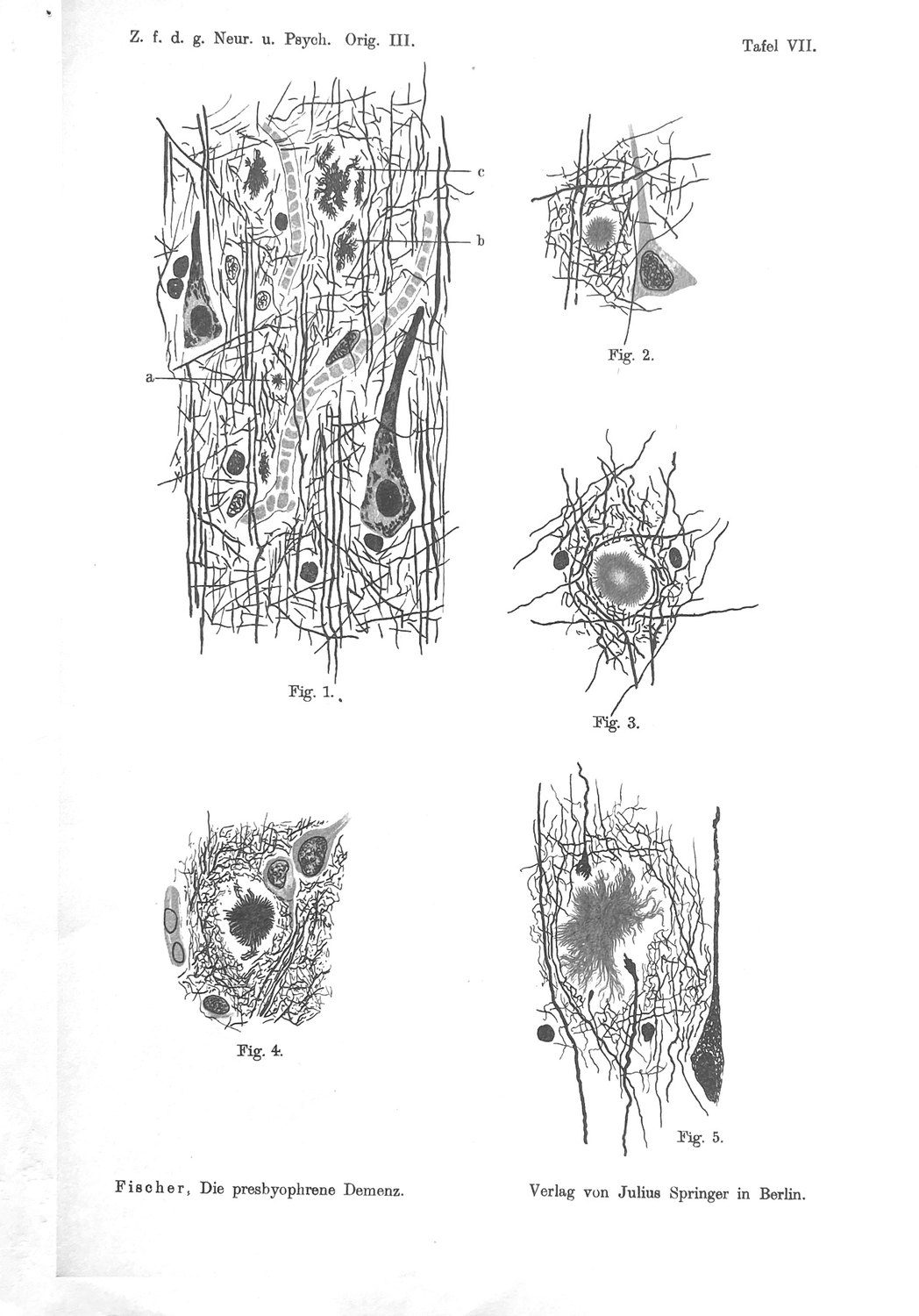
Fischer's account of Stage I is the most consequential paragraph in the monograph, and it is descriptive rather than theoretical.
"Diese kleinsten Sternchen haben meist den Durchmesser von etwa 2 µ; sie liegen bald einzeln, bald in Gruppen beisammen und konfluieren dann sehr häufig… Zugrunde gegangene Elemente oder in Destruktion befindliche Gewebsbestandteile sieht man in der nächsten Nähe der Sternchen nie; sie liegen meistens in dem nicht wesentlich veränderten Fasergewirr der Rinde, wobei die Achsencylinder um die größeren Sternchen immer, um die kleinen sehr häufig bogenförmig verlaufen, ein Verhalten, welches nur als Ausdruck einer Verdrängung des nervösen Gewebes durch die Sternchen… angesehen werden kann."
Two micrometres; lying free; confluent; and — the clause that decides several later arguments — one never sees perished elements or tissue components in destruction in their immediate vicinity. The axis cylinders bend around them. Fischer's word for what the deposit does to the tissue is Verdrängung, displacement, and he uses it consistently for the whole of Stages I–V, reserving destruction for the infiltrative mode.
Stage II adds regularity and a chemical observation.
"In diesen Drusen sind die einzelnen Fäserchen streng radiär angeordnet, verlaufen ganz gerade und sind auch meist gleich lang, so daß die Druse wie gleichmäßig zugeschoren aussieht."
Strictly radial, straight, of equal length, so that the druse looks evenly shorn. Size 8–30 µ. And:
"ein Teil dieser Drusen färbt sich nämlich wie die vorigen tiefschwarz, aber die meisten derselben nehmen eine braunviolette bis rötlichgelbe Tinktion an, wodurch sie sich sehr deutlich von dem ganz schwarz gefärbten nervösen Geflecht der Rinde abheben."
Most morning stars take a brown-violet to reddish-yellow tint that distinguishes them sharply from the black-stained nervous mesh. Fischer footnotes that his plates render this metachromasia in a single nuance for simplicity — an honest caption about the limits of the illustration. The colour is not decoration; it becomes his clock (Section XII) and his proof (Section VII).
Stage II again lies free: "Auch diese morgensternähnlichen Drusen liegen immer frei im Gewebe und verdrängen es… Nie lassen sich in der nächsten Nähe irgendwelche degenerierende oder sonstwie zugrunde gehende nervöse Bestandteile nachweisen."
The halo appears at the end of Stage II and it is a space, not a substance. Of Fig. 4: "das Gewebe ist ringsherum retrahiert, so daß ein freier Hof herum entsteht" — the tissue is retracted all around, so that a free halo arises. The Hof is what the neuropil vacates, not what the deposit adds. This is the single most misquoted feature of Fischer's description and it is unambiguous in the German.
The modern correlate, and where it fails. Stages I and II correspond to the deposit before compaction: small, without a dense core, without a neuritic reaction. But the correspondence is not exact, and the inexactness runs in Fischer's favour. Bielschowsky demonstrates fibrillar material; it is relatively blind to the loose, non-fibrillar deposit that constitutes the majority of the amyloid load in unimpaired elderly brains and that a modern immunohistochemical series finds readily. Fischer's Stage I is therefore not the first deposit but the first fibrillar deposit. There is a stage zero in front of it, invisible to him, and it is the most numerous form of all. His finding that neuritic injury is absent from Stages I and II is accordingly conservative: the still-earlier non-fibrillar forms are, on modern evidence, more inert still.
VII. Stages III and IV — The Spoke and the Wheel
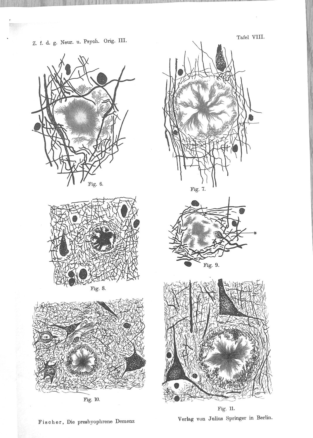
At Stage III the deposit acquires a geometry. Strands grow radially out of the morning star, the tissue retracts, and a halo opens. At Stage IV the structure is complete: a centre, radial spokes, a bounding ring. Fischer calls it a Rädchen, a little wheel, and Fig. 7 is its type specimen.
This is where the argument about what the plaque is made of was settled, in 1910, against the reading that has recently been attributed to Fischer himself. He raises the objection first:
*"So kann man bei Fig. 8 (Taf. VIII) leicht den Eindruck gewinnen, daß der das Nervengewebe umgrenzende Ring nur durch eine Verdichtung der Nervenfibrillen dargestellt wäre. Dagegen sprechen viele Momente."*
("With Fig. 8 one can easily gain the impression that the ring bounding the nervous tissue is represented merely by a condensation of the nerve fibrils. Many considerations speak against this.")
And answers it with three:
First, the colour of the whole. In Fig. 7 the central core, the strands and the marginal ring present "in gleicher Tinktion und als aus denselben Elementen aufgebaut" — in the same tint and as built out of the same elements. If the ring were host fibrils condensed, it would take the host's colour. It does not; it takes the druse's.
Second, continuity across a colour boundary. In Figs 10 and 11, where the ring is black and the strands brown, "die Fäserchen der letzteren in die ersteren übergehen oder zumindest sich mit ihnen verflechten" — the fibrils of the latter pass over into the former, or at least interweave with them. The two differently coloured parts are one structure.
Third, calibre and course. "schließlich sind alle diese Fädchen viel feiner als die Nervenfibrillen und ihr Verlauf viel wirrer und eckiger, als man es sonst bei Fibrillen zu sehen gewohnt ist." The threads are much finer than nerve fibrils and their course far more tangled and angular.
He makes the same point negatively at Fig. 16: "Nie aber ist auch nur eine Spur eines Überganges des nervösen Gewebes in die Fädchenmassen zu sehen" — never even a trace of a transition of nervous tissue into the thread masses. And he generalises it: "daß wir in diesen Drusen etwas dem Nervensystem morphologisch und chemisch ganz Fremdes vor uns haben."
The consequence for the historiography is direct. It has lately been proposed that Fischer described the plaque halo as a densification of the cortical ground substance, and that he therefore founded an extracellular-matrix theory of the disease which the amyloid era displaced. The proposal attributes to him the first impression he named in order to refute, and it discards the instrument — his own metachromasia — with which he refuted it. Whatever the merits of a matrix account of the plaque, and Section XXIV grants it several, it cannot be sourced to Fischer. He examined it and said no.
The modern correlate. Stages III and IV are the emergence of core-and-corona geometry, and this is the best-evidenced junction in the whole mapping. Condello, Yuan, Schain and Grutzendler showed by high-resolution confocal and in vivo two-photon imaging that microglial processes form a physical barrier around the deposit; that plaque microregions covered by microglia are compact and low in Aβ42 affinity; that uncovered microregions carry high-affinity protofibrillar Aβ42 hotspots; and that the hotspots are where axonal dystrophy is severe. Fischer's Hof — the space the neuropil vacates and across which his spokes run — is the compartment in which that gradient lives. He could see the geometry and not the chemistry; the modern work supplies the chemistry and inherits the geometry.
VIII. Stage V — The Thick-Fibred Skein
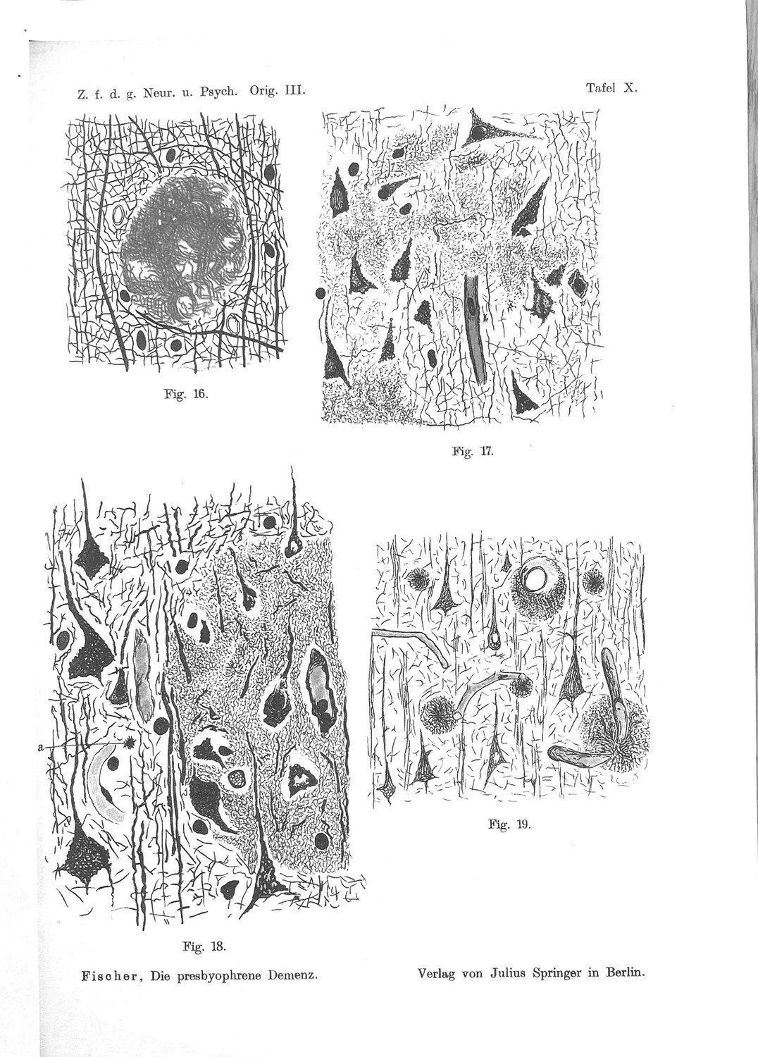
The oldest member of the growth series is the one Fischer names for its texture.
"Auch sie bestehen aus Fäserchen, die aber viel dicker sind und sich in mehr oder weniger parallel ziehenden, etwas wellig angeordneten Strähnen zu einem zierlichen Flechtwerk verbinden… sehe ich diese Formation „des dickfaserigen Knäuels" als das älteste Stadium des Prozesses an, schon deswegen, weil sie die größten Dimensionen unter allen Drusen aufweisen."
Thicker fibres, wavy parallel strands, a delicate basketwork; the largest dimensions of any druse. His argument for its being oldest is size, and he says so plainly rather than dressing it up.
Two observations attach to the mature deposit that bear on later sections.
The centre is empty of axons; the periphery is full of them. "Die periphere Zone der Drusen enthält demnach häufig ein dichtes Netz von Fibrillen, das Zentrum ist aber meistens frei von Achsencylindern und Nervenfasern, nur ausnahmsweise ziehen auch isolierte Achsencylinder noch durch." A dense fibril net at the rim, a core free of axis cylinders and nerve fibres, with only exceptional axons crossing. That is the modern dense-core plaque exactly: an acellular, afibrillar core with a corona of processes.
Cells inside drusen are rare, and Fischer says so. "die kleineren derselben sind immer zellfrei; in den größeren findet man hin und wieder Kerne resp. Kerndetritus, deren Provenienz nicht klar ist… Doch sind solche Vorkommnisse immerhin eine Seltenheit." Smaller wheel-stage drusen are always cell-free; larger ones occasionally contain nuclei or nuclear detritus, plausibly cells enclosed during growth; but such occurrences are a rarity.
That sentence disposes of a tempting modern reading, including one we have previously offered. It is attractive to suppose that the nuclei Fischer saw inside his mature deposits were the plaque-associated microglia of the modern mantle, and that he had the cells and read the direction of causation backwards. He did not. A mantle covering half the surface of a deposit is not a rarity, and Bielschowsky silver does not demonstrate microglia. The honest statement is that the cellular mantle was invisible to his method, not that he misinterpreted it — and the invisibility is itself informative, because it means everything Fischer concluded about the deposit was concluded without any knowledge of the cell that builds it.
IX. Stage VI — The Fur-Trimmed Vessel
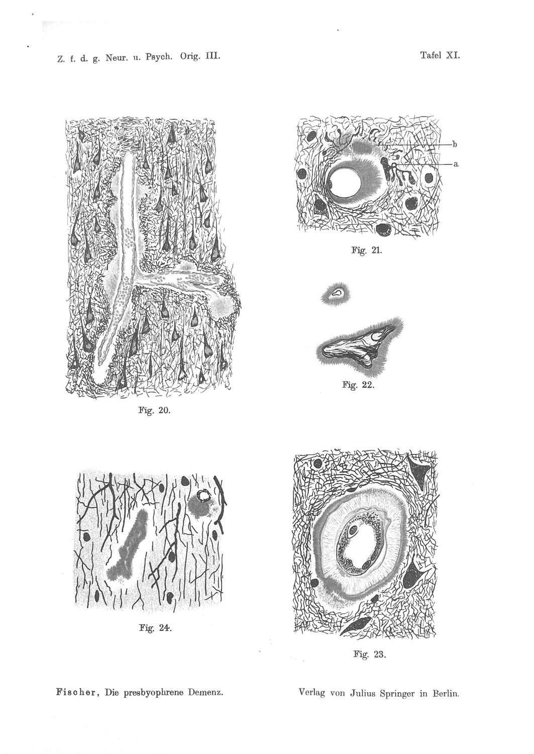
Fischer's own word for the vascular deposit is Pelzbesatz — fur trim — and it appears in his case protocols as a scored finding, including for cerebellar vessels. Figures 21 and 23 of Tafel XI are, to a modern eye, unmistakable: a vessel lumen, a wall, and a dense radial investment standing off it like a pelt.
He treats the vascular involvement as an argument, not merely an observation, and the argument is directed against the idea that the drusen are ordinary breakdown products:
"Ein Abbauprodukt, das so schön regelmäßig die Gefäße umscheidet und dabei die Wand des Gefäßes so zierlich und regelmäßig destruiert, würde etwas ganz Sonderbares darstellen."
And he adds a boundary condition of great precision:
"daß die Drusen streng mit dem Rande des Nervengewebes aufhören, daß auch diejenigen Rindengefäße, welche von den Drusen auf lange Strecken eingeschlossen werden, diese Umkleidung mit dem Moment verlieren, wie sie in die Meningen oder in die weiße Substanz eintreten."
("that the drusen stop strictly at the border of the nervous tissue; that even those cortical vessels which are enclosed by drusen over long stretches lose this investment the moment they enter the meninges or the white matter.")
Where this converges with the modern account, and where it does not. It converges on the fact: the same material occupies parenchyma and vessel wall, which is the morphological form of a molecular identity established seventy-four years later. It converges on the perivascular route, which Fischer names explicitly — "es gehört zur Regel, daß Abbauprodukte den Weg der perivaskulären Lymphräume nehmen", it is the rule that breakdown products take the way of the perivascular lymph spaces — and then argues that his drusen sit in the tissue itself despite their perivascular arrangement.
It diverges on direction and on territory. Fischer read the vessel involvement as the deposit invading and destroying the wall. The modern account of cerebral amyloid angiopathy runs the other way along the same anatomy: amyloid-β drains from the interstitium along vascular basement membranes, and deposits in the wall when that drainage fails. And modern CAA prominently involves leptomeningeal arteries, whereas Fischer's deposit stops at the pial boundary. That second divergence is worth stating rather than smoothing: either his Pelzbesatz is not co-extensive with what is now scored as CAA, or the cortical and leptomeningeal compartments differed in his material in a way that has not been revisited.
Two facts give this stage a significance out of proportion to its rank in Fischer's list. The peptide that defines the modern disease was purified from Fischer's sixth stage, not his fifth: Glenner and Wong obtained it in 1984 from cerebrovascular deposits in meningeal vessels, and Masters and colleagues from plaque cores the following year, the identity of the two establishing the continuity Fischer had asserted on morphological grounds. And Stage VI now governs the safety of the field's principal therapeutic strategy, since the amyloid-related imaging abnormalities that limit the dose of anti-amyloid antibodies arise from the vascular compartment and track the burden of CAA and APOE ε4. In Fischer's terms: the risk of attempting Stage VII is set by the state of Stage VI.
X. Stage VII — The Dissolving Druse
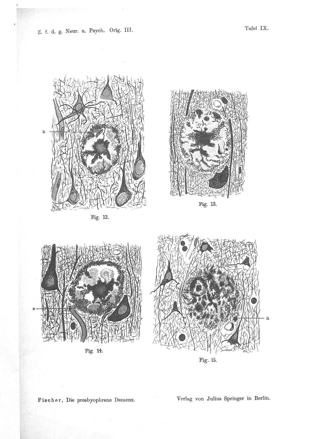
Fischer's seventh stage is the deposit coming apart. Its distinguishing feature is the Scholle — a clod or floe — and he devotes a page to deciding what the clods are.
They are not, he concludes, cell remnants, and the argument is quantitative:
"Weiter fällt es auf, daß die Schollen in den Drusen unverhältnismäßig häufiger zu sehen sind als Zellen und Zellreste; wenn nun erstere aus letzteren entstehen würden, müßte man viel mehr Übergänge sehen, als es in Wirklichkeit zutrifft."
If clods arose from cells one would see many more transitional forms than one does. His alternative:
"Das Studium einer größeren Zahl solcher Formationen läßt mir die Annahme am wahrscheinlichsten erscheinen, daß wenigstens ein Teil der Schollen durch Zerfallsprodukte der Fädchenmassen gebildet wird."
At least part of the clods are breakdown products of the thread masses themselves — the deposit digesting into its own debris. On that reading Fig. 15 is the earlier phase, threads and clods intermixed with the clods predominating, and Fig. 13 the later, where only sparse thread remnants remain at the clod margins.
And there is a chemical observation attached, easily missed: "Bei der Behandlung nach Marchi werden die in diesen Schollen eingeschlossenen Körnchen bräunlich bis schwarz." Marchi is the classical stain for degenerating myelin and for lipid. The clods are Marchi-positive. Fischer therefore reports, in 1910, that the dissolving plaque generates lipid-containing granular material — a finding whose modern echoes run from the lipid content of plaques to the lipid-droplet-laden microglia of the aged and diseased brain.
The modern correlate is the one experiment nobody could have run. Stage VII is the only one of the eight that twentieth- and twenty-first-century medicine has learned to produce deliberately. Active immunisation against Aβ42 in the AN1792 trial cleared plaques from the cortex of a subset of patients, in some cases virtually completely, and the long-term follow-up reported that clearance did not prevent progressive neurodegeneration: seven of the eight immunised patients coming to post-mortem, including those with virtually complete plaque removal, had severe end-stage dementia, with no evidence of improved survival or time to severe dementia against placebo. Fourteen-year follow-up found persistent neuropathological effects without corresponding clinical benefit.
Fischer's scheme offers three readings of that result and they compound rather than compete. The intervention arrives at the wrong stage, since by the time a patient meets criteria the population is dominated by mature deposits and the injury recorded at Stages IV and V has already happened. Most of what is removed was never doing harm, since on Fischer's own rule the neuritic reaction attaches to a minority of deposits at a particular age of the process. And dissolving a compacted deposit returns its contents to the tissue — which is exactly the transition Fischer describes at Stage VII, threads giving way to diffusely scattered granular clods.
XI. Stage VIII — The Younger Process
Stage VIII is the infiltrative mode, illustrated in Figs 17 and 18 of Tafel X above. Its description is unlike anything else in the monograph:
"hier sind an Flächen, welche manchmal auch größer als das Gesichtsfeld einer Immersion sind, die Achsencylinder und das fibrilläre Netzwerk vollkommen geschwunden und deren Stelle wird eingenommen von den erwähnten Massen… Vom nervösen Gewebe persistieren darin nur die Ganglienzellen, die sehr schwer geschrumpft sind und den größten Teil ihrer Fortsätze verloren haben."
Over areas sometimes larger than an immersion field, the axis cylinders and the fibrillar network have vanished completely and their place is taken by the thread masses; of the nervous tissue only the ganglion cells persist, severely shrunken, having lost most of their processes. Elsewhere Fischer is emphatic that his drusen displace the tissue; here, and only here, they destroy it.
The material is the same. "Ein genauerer Vergleich dieser Fädchenmassen mit den Drusen zeigt, daß sie morphologisch aus denselben Fäserchen bestehen, die den äußeren Ring der größeren Drusen und dessen Übergang in die Faserzöpfe bilden." — the infiltrating masses consist of the same fibrils that form the outer ring of the larger drusen. And transitional forms exist: Fig. 40 shows a marginal ring that has attained unusual thickness and no longer bounds itself sharply but "strahlt infiltrierend aus", radiates out infiltratingly.
What distinguishes the mode is speed against resistance. The tissue does not retract because it is not given time to:
"das infiltrative Durchwuchern, als Ausdruck einer sehr schnellen Wucherung, bei der das nervöse Gewebe keine Zeit zur Retraktion mehr hatte"
And this is why Fischer places Stage VIII outside the age series and calls it younger. The infiltrative masses stain only black — the young colour, on his clock — "was auch dafür sprechen könnte (ich betone hier das „könnte"), daß wir es hier mit Bildungen jüngeren Datums zu tun haben, d. h. mit Bildungen, die sich kürzere Zeit vor dem Tode gebildet haben." Formations of more recent date, laid down a shorter time before death. The parenthetical emphasis on could is his.
This is the most interesting stage in the scheme and the most thoroughly lost. Every account, ours included, has read VIII as end-stage confluent pathology — the natural assumption, given that it is numbered last and looks devastating. Fischer says the opposite twice: it is younger, and it advances where the tissue resists less. His eighth stage is not a measure of how long the disease has run but of the ratio between the rate of deposition and the capacity of the host to accommodate it.
The 1912 paper turns that into a clinical claim. Case 12, an acute delirium of a few days' onset, showed "Reichliche Aussaat kleinster Sterndrusen und massenhafte Infiltrate. Keine älteren Formen" — abundant sowing of the smallest star-drusen and massive infiltrates, no older forms at all — in a brain with no atrophy, minimal glial proliferation and no other parenchymal change. Fischer sets it beside Case 33 of the earlier series and draws the inference in spaced type:
"daß eine ganz akute frische Sphaerotrichieaussaat zu einem akuten Delirium führt, welches in Heilung übergehen kann; die Drusen bleiben im Gehirn bestehen, nehmen allmählich die reiferen Formen an, und wenn es dann wieder zu einem Schub einer Fädchenbildung kommt, so entsteht von neuem ein deliranter Zustand."
An acute fresh sowing produces a delirium that can remit; the drusen persist and mature; a further Schub — a wave, a relapse — of thread formation produces a new delirious state. That is a model of episodic deposition with maturation between waves, proposed on the strength of two cases and offered as such.
XII. The Colour Clock
Running underneath the stage table is an instrument, and it is the most original thing in the monograph.
"Wir sehen nämlich, daß die Sternform als die jüngste Bildung immer nur schwarz gefärbt erscheint, wogegen die Morgensterne und die Strähnchen sehr häufig eine bräunliche Färbung annehmen; wir könnten also daraus auch schließen, daß sich die Fädchen in den jüngsten Stadien schwarz, in den späteren braun färben."
Threads impregnate black when young and brown when older. Fischer then applies the rule within a single deposit:
"Das gleiche Verhalten bemerkt man nun auch im Stadium der Rädchenbildung; die zentralen Partien sind daselbst meist bräunlich oder rötlich tingiert, wogegen der Randring immer schwarz erscheint… daraus folgt, daß auch auf Grund des tinktoriellen Verhaltens der Randring als jüngste Bildung der Drusen erscheint."
In a wheel-stage druse the centre is brown and the rim is black. If black is young, the rim is the most recently formed part. Therefore the deposit grows outward from the centre, adding new material at its margin.
That is a growth model derived from a staining property, tested on a second observable — the size series — and consistent with it. It is also, so far as we can establish, the first proposal that a plaque grows by peripheral accretion, and it agrees with what in vivo two-photon imaging now shows directly: plaques appear, and then enlarge outward from an established core.
The inference has a limitation Fischer does not state and a reader should. Silver impregnation intensity depends on many things besides the age of the substrate — section depth, fixation, local pH, the density of the material — and a colour difference between core and rim could reflect packing rather than chronology. Fischer's defence is indirect but real: the same colour ordering holds across deposits of different sizes as it does within one, and two independent orderings agreeing is better than either alone.
He uses the same instrument once more, and it is the point at which the clock and the stage table meet. Because the infiltrative masses of Stage VIII stain only black, he concludes they are young — which is why the eighth entry in a list ordered by age is nonetheless the second-youngest thing in it.
Part Three — The Rules Fischer Derived
XIII. The Neurite Rule
The most consequential sentence Fischer wrote is on page 381 of the 1910 monograph, and it has never been quoted whole.
"Die kolbigen Wucherungen der Achsencylinder kommen nur in etwa 50% der Fälle vor; dabei auch nur um die größeren Drusen; am häufigsten finden sie sich um das Stadium V, dann um Stadium IV, seltener um Stadium III, nie in der Nähe der Stadien I und II, ebenso auch nie um Stadium VIII. (Siehe Einteilung der Stadien auf Seite 392.) Warum sich in den einzelnen Fällen die Keulen vorfinden, in den anderen nicht, ist nicht klar geworden, im histologischen Verhalten war sonst keine Differenz merkbar."
("The club-shaped proliferations of the axis cylinders occur in only about 50% of cases; and then only around the larger drusen; most frequently they are found around Stage V, then Stage IV, more rarely around Stage III, never in the vicinity of Stages I and II, and likewise never around Stage VIII. (See the classification of stages on page 392.) Why the clubs are present in individual cases and not in others has not become clear; in histological behaviour no other difference was perceptible.")
Four things in that sentence deserve separate attention.
It is a per-lesion measurement with the brain as its own control. Every druse in a section is an independent observation scored for two variables — its stage and whether clubs surround it — in the same tissue, the same subject, the same fixation, the same stain. Age, agonal state, post-mortem interval, disease duration and genotype are held constant by construction. This is the design quantitative neuropathology now regards as the strongest available for questions of local pathology, and Fischer used it because it was the only way to extract an ordering from static material.
The relation is an inverted U. Clubs peak at Stage V, decline through IV to III, are absent at I and II — and are absent again at VIII. No secondary account reports the second half of that. The clause "ebenso auch nie um Stadium VIII" is not in Goedert's summary and it is not in any treatment of Fischer we have found. It changes the shape of the claim from a ramp to a curve, and the curve has a mechanism attached: Stage VIII destroys the axons rather than swelling them. Fischer notes in the same section that within the infiltrated areas only the thicker traversing axis cylinders take the stain at all, "wogegen die größte Mehrzahl derselben ungefärbt, aber bei stärkerer Abblendung noch deutlich sichtbar ist" — the great majority unstained though still visible under strong stopping-down. There is no club because there is no longer an axon in a condition to make one.
The "50%" attaches to cases, not certainly to plaques. "der Fälle" in Fischer's German most naturally means cases — patients — and the per-deposit restriction is carried by the clause that follows, "dabei auch nur um die größeren Drusen." Goedert renders the figure as fifty per cent of plaques. The German will bear either reading and we do not resolve it, because the argument does not need it: the stage-ordering is unambiguous whichever way the percentage is taken, and it is the ordering that does the work. What can be said without ambiguity is that in half of Fischer's cases the deposit provoked no visible neuritic reaction anywhere, and that where it did, it did so only around the older and larger forms.
Fischer marks the residual as unexplained. "Warum sich in den einzelnen Fällen die Keulen vorfinden, in den anderen nicht, ist nicht klar geworden." He has a variable that explains part of the variance and he says so, and then says the rest is unaccounted for. That is the correct report, and it is the sentence that separates his staging from a taxonomy: he expected the scheme to predict something, checked whether it did, and recorded the shortfall.
The clubs themselves are described with care. Axis cylinders near drusen show spindle-shaped swellings ranging from "ganz leichte, manchmal gerade noch merkbare Verdickungen" to "eine Mächtigkeit, die der Größe der Ganglienzellen gleichkommt" — from barely perceptible thickenings to swellings equalling a ganglion cell in bulk. A single axon may carry one or several. Clubs may sit on an already spindle-swollen axon. And they are arranged around the deposit: one of his figure captions reads "Kranzartige Anordnung der kleinen Keulen" — wreath-like arrangement of the small clubs.
Fischer's five-part definition of the process, quoted in Section V, states the rule as a defining property rather than an incidental finding: the formations are those which lead to proliferations of the axis cylinders "in einem kleinen Prozentsatz der Fälle und in bestimmtem Alter des Prozesses" — in a small percentage of cases and at a definite age of the process. The stage-conditionality of injury is written into his definition of what the lesion is.
XIV. Whose Axons? The Parent Cell of the Club
A modern reader brings to Fischer's clubs a question he could not have known would be asked: does the wreath of dystrophic neurites around a deposit belong to one neuron, or to many? The answer decides what kind of object the plaque is. If the corona were a single cell's arbor, the deposit would be a monument raised where a neuron was dying, and the plaque count would be a proxy for cell death. If instead it is the traffic of the neuropil — fibres arriving from elsewhere, most of whose cell bodies are nowhere near — then the plaque is a lesion of the wiring that passes through it, and it can injure far more neurons than it stands beside.
Fischer answered the question, at length, on pages 379 to 381, and no account of him quotes the passage. His answer is: many, and from outside.
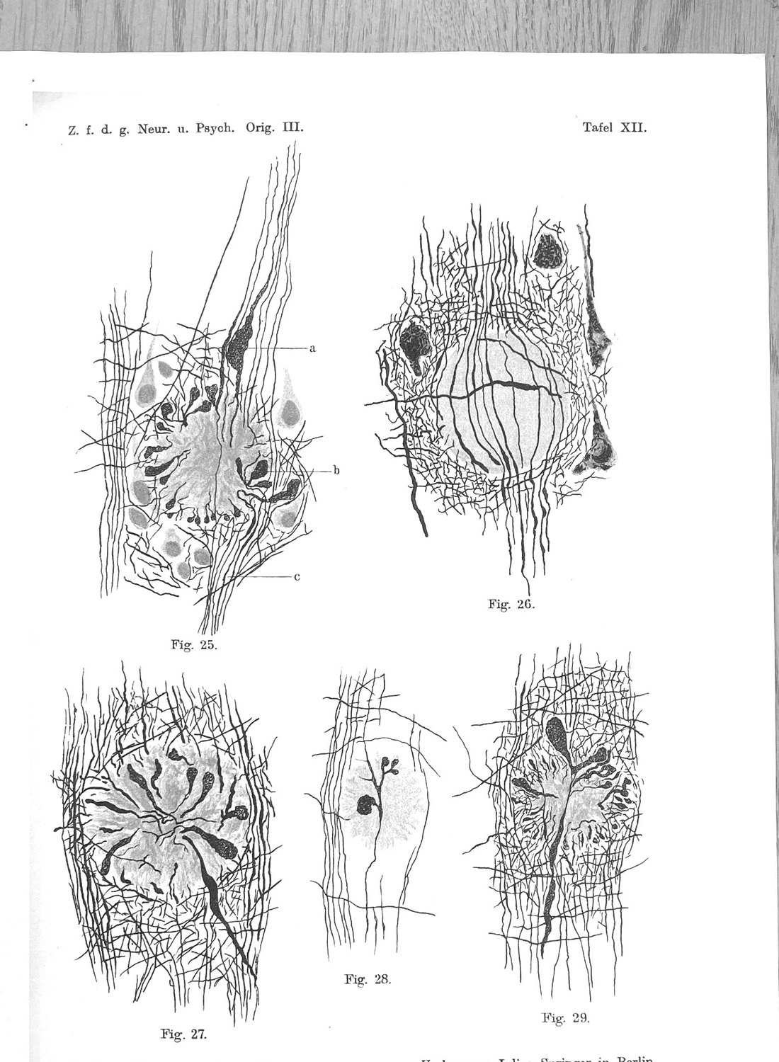
He traced the fibres, and he says how. "Bei einer großen Zahl dieser Kolben kann man einen direkten Zusammenhang des fädigen, zentral gerichteten Endes mit den Achsencylindern der Umgebung nachweisen, was um so häufiger gefunden werden kann, je dickere Schnitte man untersucht." — in a large number of these clubs a direct continuity can be demonstrated between the thread-like, centrally directed end and the axis cylinders of the surroundings, and the thicker the sections examined, the more often it can be found. The dependence on section thickness is the tell: the parent fibre is usually out of the plane of the cut, which is to say it comes from somewhere else. He states the control in the next sentence — "An diesen sind aus selbstverständlichen Gründen auch die Achsencylinder auf größere Strecken zu verfolgen, so daß… eine Verwechslung mit anderen Elementen ausgeschlossen werden kann" — in thick preparations the axis cylinders can be followed over longer stretches, so that confusion with other elements is excluded.
The clubs point outward and overrun the deposit. They are "immer kranzartig um die Druse gestellt, und zwar so, daß der dünne Hals nach innen steht, das kolbige Ende nach außen gerichtet ist und das Areal der Druse überschreitet" — always set wreath-like about the druse, thin neck inward, club end directed outward and exceeding the area of the druse. A swelling whose bulk lies outside the lesion is not a structure the lesion produced. It is a structure the lesion caught.
Figure 25 is drawn as separate fibres, and Fischer reads it that way. The fibril at c winds through several vertical bends that the drawing cannot render, thickens at one point, enters the druse, turns in a sharp arc, passes over into a large club and there ends. "Ein anderer Achsencylinder" — another axis cylinder — likewise reaches the margin after several windings and ends "in kleeblattartiger Dreiteilung mit drei Keulen (bei b)", in a cloverleaf trifurcation with three clubs, at b. Two fibres, two courses, two letters on the plate, and a third proliferation keyed at a. Nothing in the figure is drawn as one arbor.
Most clubs are not endings at all. This is the observation that settles the question. "Häufig zeigt sich, daß die Keulen dort, wo man sie und ihre Achsencylinder auf längere Strecken verfolgen kann, nicht als Endkeulen eines Achsencylinders aufsitzen, sondern als seitlicher Auswuchs sich darstellen." — frequently, where the clubs and their axis cylinders can be followed over longer stretches, they do not sit as terminal clubs of an axis cylinder but present as a lateral outgrowth; one such axon carries either a single outgrowth or several. And he names the figure that demonstrates it: "Besonders instruktiv und einwandfrei zeigt sich das Verhalten der Keulen zu dem Achsencylinder in der Fig. 28 (Taf. XII), welche einen Tangentialschnitt einer Druse darstellt; darin ist ein Achsencylinder, der über der Druse verläuft und gerade in deren Randpartie zu liegen kommt, in ganzer Länge getroffen und zeigt mehrere Auswüchse." A tangential section catches, along its whole length, an axis cylinder that runs over the deposit and grazes its margin, and that one fibre bears several clubs. It arrives from outside the field, is injured in passing, and continues.
He separates the fibre populations by their course. The axis cylinders in the immediate surroundings are "durchwegs zur Seite gedrängt, was am deutlichsten an den bündelweise verlaufenden Radiärfasern zu sehen ist" — pushed aside throughout, most clearly among the radial fibres running in bundles; whereas "die unregelmäßig und quer verlaufenden Fasern der Rinde endigen scheinbar am Rande der Drusen, in Wirklichkeit aber winden sie sich mehrfach hin und her und häufig enden sie dann in einer Keule." The irregular, transversely running cortical fibres only appear to stop at the rim; in fact they wind back and forth and often then end in a club. Radial bundles and tangential fibres are different traffic with different origins, and both are represented in the same wreath.
The neurons standing nearest are not the sick ones. Fischer scored the ganglion cells separately and found every grade of change, from the faintest to the most severe. Then he stated where the change is, on pages 381 to 382: the uppermost cell layers are always the most heavily affected, and "die Veränderung [beschränkt sich] nicht auf die den Drusen am nächsten liegenden Zellen…, sondern [ist] mehr diffus" — the alteration is not confined to the cells lying closest to the drusen but is more diffuse. A local parent cell in collapse, feeding a corona of its own dying processes, is the picture his own cell scoring excludes.
What his stains establish, and what they cannot. Two dissociations carry the identification, and both are on page 383. Cajal's method "färbt die Drusen nur sehr undeutlich, wogegen die Keulen genau so wie die Achsencylinder tief schwarz gefärbt sind" — stains the drusen only very indistinctly, while the clubs stain deep black exactly as the axis cylinders do. Weigert's glial method leaves the drusen wholly uncoloured, and "auch die Keulen bleiben ganz ungefärbt, auch dann, wenn man sonst tadellose Ausfärbung der Gliafasern erzielte" — the clubs too remain unstained, even when the glial fibres take the stain faultlessly. The club follows the axon under a neurofibrillary method and refuses a glial one: it is axoplasm, neither deposit nor glia. What no method of 1910 could do is type the parent cell, and Fischer never claims to have done it. He writes Achsencylinder der Umgebung and stops, which is the correct stopping place — and the reason the proposal that his clubs are the dystrophic processes of parvalbumin-expressing interneurons cannot be sourced to him (Section XXVI). That proposal is not merely unsupported by his text; the en passant morphology points away from it, since a swelling on the flank of a fibre in transit is silent about whose fibre it is.
The modern record has answered the question he framed. Adalbert and colleagues traced axons longitudinally through, distal to and proximal from plaque-associated dystrophies in a transgenic model and found that the axons remained continuous and that the corresponding neurons not only survived but remained morphologically unaltered — the dystrophy is a local swelling on a through-running fibre whose cell body is elsewhere and metabolically intact. Yuan and Grutzendler then showed that these plaque-associated spheroids form on long-range axons and act as size-dependent current sinks producing action-potential conduction blockade, so that the functional lesion is to connectivity passing through the deposit rather than to a neuron residing at it. And the corona is enriched for presynaptic elements: Sadleir and colleagues characterise the dystrophies as presynaptic, disrupted in their microtubules, elevated in BACE1 and generating Aβ locally. The wreath around a single plaque is contributed by many neurons, most of whose cell bodies are neither local nor dying — which is what Fischer reported, in the vocabulary of the bent, swollen, passing axis cylinder.
Two consequences follow for the rest of this paper. The first is a caution about reading his plates. The radial spokes of Stages III and IV are the druse's own threads and are not neurites at all; Fischer separated them from the axons by tint and by the metachromasia of his stain (Section VII), and a modern eye that takes the spokes for a converging arbor has mistaken the deposit for its victim. The second is a confound that the operational index of Section XXVII inherits. If neuritic dystrophy is a property of the fibres that pass a deposit, then dystrophy per plaque is in part a function of local fibre density and orientation, and two deposits sitting in tissue of different wiring density are not comparable on that term alone. Fischer supplies the correction himself, by scoring the radial bundles and the tangential fibres as different populations. A modern index should do at least as much.
XV. Displacement, Not Destruction
Fischer's second rule is a distinction he maintains across the whole monograph and which the word plaque has since erased.
The drusen of Stages I–V displace the tissue. He establishes it from the smallest form onward, from the behaviour of the axons: "die Achsencylinder um die größeren Sternchen immer, um die kleinen sehr häufig bogenförmig verlaufen, ein Verhalten, welches nur als Ausdruck einer Verdrängung des nervösen Gewebes durch die Sternchen… angesehen werden kann." Axis cylinders bend around the deposits. That is a mechanical relation, and it is what the Hof records: tissue that has moved aside.
They do not, in the main, destroy it. His summary definition puts it in the conditional: the formations "das Gewebe zwar schädigen, es aber nur ausnahmsweise schwerer destruieren, und zwar dann, wenn sie es diffus infiltrieren oder einschließen." They damage the tissue but destroy it more severely only exceptionally, namely when they diffusely infiltrate or enclose it.
This is why Stage VIII sits outside the series, and it is also the single point on which Alzheimer and Fischer agreed. Alzheimer used the observation against him — plaques displace rather than destroy, therefore they cannot be the cause of the dementia — and Fischer accepted the premise while rejecting the conclusion. Section XXII takes up that exchange.
The distinction has a modern descendant that neither man could have drawn. What injures the neuropil around a modern plaque is not the volume the deposit occupies but the gradient of diffusible species it maintains at its edge, and the extent of that gradient depends on whether the deposit is compacted and covered. A deposit that displaces without destroying is, in modern terms, a deposit whose halo is short. Fischer had the phenomenon, expressed as a mechanical fact about bent axons and a vacated space, and no access to the chemistry that makes the space dangerous or safe.
XVI. No Reactive Inflammation — and What Fischer Did With It
The fifth of Fischer's defining properties is a negative: the formations "keinerlei reaktive Entzündung hervorrufen", call forth no reactive inflammation whatever.
That clause has been quoted in recent work as though Fischer were reporting a quiet, non-inflammatory remodelling of the tissue. He was not. He was killing a hypothesis, and the hypothesis was his own.
Fischer took seriously — more seriously than any account of him admits — the possibility that the drusen were an organism. The morphological case is stated at length: "die Drusen beinahe in allen ihren Stadien eine große morphologische Ähnlichkeit mit Fadenpilzen haben", and he lists what a living, organised matter would explain that a dead deposit would not:
"Der Aufbau aus Fädchen, die verschiedene Stellung, Färbung und Dicke bei verschiedenem Alter, das scheinbar aktive Wachstum mit dem konsekutiven Verdrängen des Nervengewebes, das infiltrative Durchwuchern, als Ausdruck einer sehr schnellen Wucherung, bei der das nervöse Gewebe keine Zeit zur Retraktion mehr hatte, und schließlich der enge Anschluß an die Gefäße und das Durchwuchern der Gefäßwand."
Build-up from threads; position, colour and thickness varying with age; apparently active growth with consequent displacement; infiltrative overgrowth as very rapid proliferation; close attachment to vessels and growth through the vessel wall. It is a good case, honestly made, for a conclusion he then refuses:
"Aber! — wo bleibt dann die entzündliche Reaktion? In unserem Falle gibt es keine Entzündung um die Drusen; die einzige Reaktion, die das nervöse Gewebe zeigt, sind die kolbigen Wucherungen der Achsencylinder, welche jedenfalls als Ausdruck einer Schädigung des Gewebes durch die fremden Einlagerungen angesehen werden müssen. Aber Pilzwucherungen, welche keine entzündliche Reaktion setzen, sind bisher unbekannt."
("But! — where then is the inflammatory reaction? In our case there is no inflammation around the drusen; the only reaction the nervous tissue shows are the club-shaped proliferations of the axis cylinders, which must in any case be regarded as an expression of damage to the tissue by the foreign inclusions. But fungal growths that set up no inflammatory reaction are hitherto unknown.")
Two points follow, and both matter for how Fischer is used.
The absence of inflammation is an argument against infection, not a characterisation of the lesion's biology. Fischer deploys it exactly once, in exactly that role. Extracting the clause and offering it as evidence that the plaque is a cold, non-inflammatory remodelling of host material inverts its function in the text — and does so in a sentence where Fischer, in the same breath, calls the deposit a foreign inclusion and the neurites damage caused by it.
And two years later he reported a glial reaction — stage-conditionally. In 1912, having once again rejected a glial origin for the deposits, Fischer records: "außerdem fanden sich besonders um die größeren und älteren Herde gewucherte und vergrößerte Gliazellen" — in addition there were found, especially around the larger and older foci, proliferated and enlarged glial cells. The qualifier is the finding. Not that glia surround the deposits, but that they surround the larger and older ones: the same conditionality he had established for the neurites in 1910, transferred to a different cell and stated as a positive observation rather than as an absence. It is also, on the modern measurement, right. In the forty-subject series of Section XIX, GFAP-positive reactive astrocytes rise with symptom duration at Kendall τ = 0.30 while microglial number does not move at all (τ = 0.045). Fischer's proliferated and enlarged glia around the larger and older foci is the astroglial arm of that result, reported on the correct conditional in 1912.
Entzündung and gliosis are different objects, and he was not confused about which one he had. The exudative, leucocytic reaction whose absence refutes a fungus is not the hypertrophy of a resident cell, and he reports the second without embarrassment in the same body of work that denies the first. What was genuinely invisible to him is the third cell: Bielschowsky silver does not demonstrate microglia. The astroglial reaction was available through the neuroglia stains in his panel; the microglial mantle was not, and his statement that nuclei inside deposits are rare is therefore a statement about a population from which the most numerous plaque-associated cell type has been silently removed.
The rejection of the microbial hypothesis was far more rigorous than "negative bacterial stains." The usual account of Fischer credits him with Gram and Ziehl-Neelsen preparations. In fact he ran four independent lines:
"Ich habe deswegen bei mehreren Fällen auch die meisten Organe des Körpers histologisch durchsucht, aber nichts gefunden, was an die Drusen erinnert hätte… Kulturversuche könnten in dieser Richtung mehr Aufschluß geben; dieselben wurden in der mannigfaltigsten Variation versucht, blieben aber ganz ohne Resultat. Auch wurden mit den die Drusen enthaltenden Gehirnen Komplementbindungsversuche angestellt, aber ebenfalls mit negativem Erfolg."
Bacterial stains; systematic histology of most organs of the body in several cases; culture in manifold variations; and complement-fixation serology on drusen-bearing brains. All negative. And an honest caveat that the organ survey proves nothing, because there is no specific stain for the threads as such — they are recognisable only by their grouping, that is, only once they are already drusen.
In 1912 he restated the disclaimer in his own defence, because the comparison had been read as a claim:
"machte aber darauf aufmerksam, daß sie in vieler Hinsicht an Streptotricheenkolonien erinnern (aber — und das möchte ich wieder betonen, — ohne dieselben je als tatsächliche Bakterien hingestellt zu haben)"
The morphological simile was a simile. Whatever else Fischer was, he was not a precursor of the infectious hypothesis of Alzheimer's disease, and the passages enlisted to make him one are the passages in which he tested that hypothesis and reported it dead.
XVII. Stage Against Duration
In 1912 Fischer did the experiment that turns his staging from a description into a claim about time. He had, by then, both morphological stages and clinical histories, and he crossed them.
"Es ließ sich nun weiter die interessante Tatsache feststellen, daß sich eine Übereinstimmung zwischen der Dauer der Krankheit und der Art der Drusen zeigte. Schon aus dem Aussehen der Drusen hatte ich geschlossen, daß die verschiedenen Formen derselben auch verschieden alten Stadien entsprechen dürften; in Übereinstimmung damit ergab dann die klinische Zusammenstellung, daß die Fälle mit kurzer Krankheitsdauer auch vornehmlich die jüngeren Stadien, jene mit längerer Krankheitsdauer die älteren Stadien der Drusen aufwiesen."
("It could further be established as an interesting fact that a correspondence appeared between the duration of the illness and the kind of drusen. From the appearance of the drusen I had already concluded that their different forms should correspond to stages of different age; in agreement with this, the clinical compilation then showed that the cases with a short duration of illness exhibited predominantly the younger stages, those with a longer duration the older stages of the drusen.")
The logic deserves to be spelled out because it is better than it looks. Fischer's ordering of the stages was derived internally, from morphology and from the colour clock — that is, from a source entirely independent of the clinical record. He then tested the ordering against an external variable, disease duration, which he had not used to construct it. The agreement is therefore a genuine validation and not a circularity, and it is the only external validation of the scheme that Fischer himself was in a position to perform.
He adds a second cross-check at the other end of the range: "Weiter ergab sich bei den deliranten Fällen, die ja als die akuteste Form aufgefaßt werden müssen, der schwerste Grad der Hirnschädigung durch die Drusen in Form der infiltrativen Wucherung der Fädchenmassen." The delirious cases — the most acute form — show the infiltrative mode. Short course, young stages, infiltration; long course, old stages. Both ends behave.
Section XIX sets this beside its modern repetition.
XVIII. Latency, Threshold, and the Two Brains That Should Not Have Existed
The 1912 paper contains a passage of general pathology that has no business being as modern as it is. Its occasion is an anomaly: of 275 cases, the proposed scheme fitted all but two — two patients who in life showed only a simple dementia without presbyophrenic symptoms and who nonetheless had drusen in the brain.
Fischer does not discard them. He notes first that the sowing of drusen in those two brains was very sparse compared with the others, and then reasons:
"ergab sich die Auffassung, daß sich hier eine Art latenter Gehirnveränderung entwickelt hatte, die erst dann zu klinischen Zeichen geführt hätte, wenn sie zu stärkerer Entwicklung gekommen wäre."
("the view emerged that a kind of latent brain change had developed here, which would have led to clinical signs only once it had come to stronger development.")
And then generalises it:
"Denn es ist wohl selbstverständlich, daß, wenn irgend einem klinischen Symptomenbild eine bestimmte Organerkrankung zugrunde liegt, dieselbe erstens bereits eine Zeit lang bestehen muß, bevor die klinischen Symptome sich entwickeln, und zweitens ein bestimmter Grad der anatomischen Laesion notwendig ist; denn die Organlaesion geht dem klinischen Symptomenbild immer unbedingt voran."
("For it is surely self-evident that when a definite organ disease underlies some clinical picture, that disease must first have existed for some time before the clinical symptoms develop, and second, a definite degree of the anatomical lesion is necessary; for the organ lesion always unconditionally precedes the clinical picture.")
A latent period and a threshold of severity, stated as the general form of the relation between a lesion and a syndrome, in 1912, and derived from two discrepant cases rather than from theory. It is the structure of preclinical disease. Fischer applies it to explain not only the two anomalies but the general phenomenon: "Deswegen ist es wohl einleuchtend, daß sich Fälle finden werden, bei denen eine Organerkrankung ohne das erwartete klinische Symptomenbild vorgefunden wird, besonders dann, wenn die Organerkrankung nur eine geringe Intensität erreicht hat (was ja wohl auch individuellen Schwankungen unterliegen dürfte)." — especially when the organ disease has reached only a low intensity, which is itself presumably subject to individual variation.
Individual variation in the threshold at which a given burden of pathology produces a syndrome is, in current vocabulary, cognitive reserve. Fischer names it in a parenthesis.
It is worth being exact about what this does and does not anticipate. It does not anticipate the modern finding, which is that a large fraction of unimpaired elderly carry substantial pathology; Fischer's rate was six per cent and his two anomalies were sparse-deposit cases. What it anticipates is the form of the explanation the modern finding requires — that the lesion precedes the syndrome by an interval, that a quantity must be crossed, and that where the quantity sits varies between people. Given that he arrived at it from two cases that did not fit, and published them, the passage is a better piece of scientific conduct than most of what has been written about him.
Part Four — Against the Modern Science
XIX. Stage Against Duration, Measured Again
Fischer's 1912 cross-tabulation of morphological stage against disease duration was repeated in 2016, with numbers, by people who had not read it.
Serrano-Pozo, Betensky, Frosch and Hyman examined dense-core plaques in temporal neocortex from forty subjects with Alzheimer's disease whose symptom durations ranged from four to twenty years, together with nine controls. They set out from the observation that plaque burden is essentially stationary across the clinical course, and asked whether the local features of individual plaques change even though their number does not.
They do. Dystrophic neurites, labelled with SMI312, rose with symptom duration (Kendall τ = 0.34, P = 0.001). GFAP-positive reactive astrocytes rose (τ = 0.30, P = 0.003). CD68-positive microglia rose steeply (τ = 0.48, P < 0.0001). IBA1-positive microglia did not move at all (τ = 0.045, P = 0.655).
The correspondence with 1912 is close enough to be worth stating precisely. Fischer: short duration carries the younger stages, long duration the older. Serrano-Pozo: the count is flat and the per-plaque injury climbs across the whole observable range of the disease. These are the same claim at different resolutions — that what accumulates over two decades of clinical course is not more deposits but more advanced deposits.
The IBA1/CD68 dissociation adds something Fischer could not have seen and which bears on his Stage V. The number of microglia at the plaque does not change across twenty years of disease; what changes is their lysosomal and phagocytic activation. The cellular census of the structure is constant and its activity escalates. Fischer's mature druse is not a static object that accumulates cells; on the modern measurement it is a structure of stable composition whose state changes — which is exactly the kind of variable a staging scheme is built to capture and a count is not.
XX. The Core and the Corona
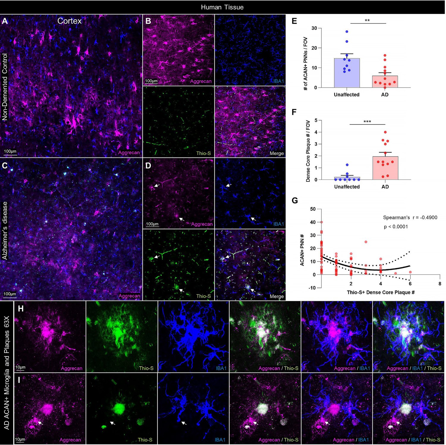
Set that panel beside Tafel VIII of Section VII and the continuity is not rhetorical. The object in the middle of both is a compact, radially organised deposit with a distinguishable centre, a bounded margin, and a zone around it in which the tissue is not as it is elsewhere. Fischer drew it in 1910 from a silver impregnation; the modern figure resolves it with three fluorophores and adds the cell he could not stain.
The mechanism that fills in his geometry was established by Condello, Yuan, Schain and Grutzendler. Using high-resolution confocal and in vivo two-photon imaging in amyloid-depositing mice, they showed that microglial processes form a physical barrier around the deposit; that plaque microregions covered by microglia are compact and show low affinity for Aβ42; that uncovered microregions show high Aβ42 affinity and are where protofibrillar hotspots form; and that those hotspots are where axonal dystrophy is severe. The barrier prevents outward expansion and holds the neurotoxic species off the surrounding neuropil.
Read against Fischer, this is an account of what determines the extent of the Hof and what happens in it. He observed that the tissue retracts and that a space opens; that spokes cross the space; that clubs appear at the margin of the larger deposits and not the smaller. The modern work says the space contains a concentration gradient, that the gradient's reach is set by how completely the deposit is covered, and that the clubs are what the gradient does to the processes crossing it.
The human demonstration is the one that decides between counting and staging. Yuan and colleagues examined Trem2- and Dap12-haplodeficient mice and, critically, post-mortem tissue from human carriers of the TREM2 R47H variant. Microglia in those tissues showed a markedly reduced ability to envelop deposits. The consequences were stage-shaped: an increase in less compact, filamentous deposits, and a greater extent of dystrophic axons and neuronal processes bearing hyperphosphorylated tau.
In Fischer's vocabulary, the R47H brain is arrested between Stages II and IV. Its deposits fail to complete the transition to the compacted wheel; the neurites around them suffer; and the plaque count is not what distinguishes it. A genotype that raises the risk of Alzheimer's disease roughly three- to four-and-a-half-fold does so by moving a morphological variable while leaving the tallied one alone — which is a fair summary of why the tally has never worked.
It is worth noting one inversion that Fischer's scheme, taken naively, gets wrong and that the modern work corrects. Because his series runs from small and simple to large and complex, and because injury rises along it, the natural reading is that compaction is deterioration. It is not. Compaction is what the tissue achieves; the filamentous, uncontained deposit is what remains when the response fails. Fischer's Stages III to V are a description of a deposit being brought under control, and the clubs that appear alongside them are the cost of a containment that is under way and incomplete. His Stage VIII — fast, uncontained, in tissue that resists less — is the failure mode, and it is the one that carries no clubs at all because it does not leave enough axon to swell.
XXI. What Is Inside the Halo
The question of what occupies the space around the deposit is the one on which Fischer has most often been enlisted, and it needs to be answered in two parts, because the honest answer is not the same in both.
What Fischer said. He said the ring is made of the same threads as the core, and he proved it with colour (Section VII). He said the halo is a retraction space. He said the deposit is "dem Nervensystem morphologisch und chemisch ganz Fremdes." He said the smallest forms lie in tissue showing no other change. On the specific question — is the plaque a rearrangement of host material? — his answer is no, given four ways.
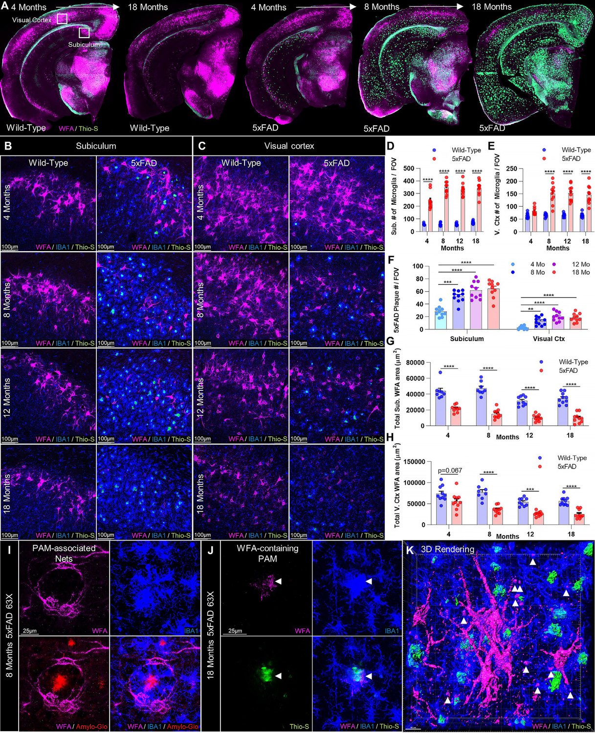
What is nonetheless true about the matrix. Crapser and colleagues report that aggrecan, the principal proteoglycan of the perineuronal matrix, is deposited within human dense-core plaques, colocalising with thioflavin-S signal; that perineuronal net material is found inside microglia in both mouse and human tissue; that net number correlates inversely with plaque count in human prefrontal cortex (Spearman r = −0.49, P < 0.0001); and that depleting microglia with a CSF1R inhibitor prevents net loss in 5xFAD mice despite the persistence of plaques. Beyond the perineuronal matrix, heparan sulfate is the most frequently encountered glycosaminoglycan in amyloid deposits, and perlecan, agrin, glypicans and syndecans are all present in plaques.
These two sets of statements are compatible, and the way they are compatible is the point. Matrix material ends up in the plaque. It does not get there by condensing in place; it gets there by being degraded elsewhere and incorporated — Crapser's own conclusion is that microglia "facilitate plaque-dependent PNN loss", with the plaque upstream. The distinction is between a rearrangement and a transport, and only the second is consistent with the cellular mechanism and with what Fischer could see.
So the plaque is in part a matrisomal structure, and Fischer was still right that it is not condensed host ground substance. The corpus's earlier reading conflated two different matrices — the basement-membrane and interstitial matrisome that is genuinely incorporated into deposits, and the aggrecan-based perineuronal lattice that is a property of particular neurons — and then attributed the composite to Fischer, who described neither. Section XXVI returns to this.
XXII. Alzheimer's Objection
The 1912 paper preserves something that exists nowhere else: Alzheimer's argument against the pathogenic significance of plaques, quoted at length by the man he was arguing with.
*"Es gibt Fälle von zweifelloser Dementia senilis, bei denen die Drusen nicht sehr zahlreich sind, außerdem verdrängen die Drusen, wie Fischer selbst betont, die nervösen Strukturen mehr, als sie dieselben zugrunde richten. So kann in diesen Fällen die Schädigung des Rindengewebes durch die Drusen keine sehr beträchtliche sein. Ferner treffen wir auch an Stellen, wo wir keine Drusen in der Hirnrinde fanden, die bekannten ausgebreiteten senilen Veränderungen… Wir fanden Veränderungen in den basalen Ganglien, der Medulla, dem Kleinhirn und dem Rückenmark, obwohl dort überhaupt keine Drusen oder nur sehr vereinzelt zu finden sind. So müssen wir doch wohl zum Schlusse kommen, daß die Drusen nicht die Ursache der senilen Demenz, sondern nur eine Begleiterscheinung der senilen Involution des Zentralnervensystems sind."*
("There are cases of undoubted dementia senilis in which the drusen are not very numerous; moreover the drusen, as Fischer himself emphasises, displace the nervous structures more than they destroy them… We found changes in the basal ganglia, the medulla, the cerebellum and the spinal cord, although no drusen at all, or only very isolated ones, are to be found there. So we must surely come to the conclusion that the drusen are not the cause of senile dementia but only an accompanying phenomenon of the senile involution of the central nervous system.")
That is the amyloid-sceptic argument in its original form, and its author is Alzheimer. Its three limbs are the ones still in use: the quantitative dissociation (dementia with few plaques), the mechanistic implausibility (displacement is not destruction), and the anatomical mismatch (degeneration where there are no plaques). It was made in 1911, against Fischer, by the man whose name the disease carries.
Fischer's reply is instructive in two directions. On the substance he concedes less than he might, because he holds that presbyophrenic dementia and simple senile dementia are two different diseases, so that cases of dementia senilis with few plaques are not counter-examples to his claim but instances of the other condition: "ich habe in meiner Publikation nie behauptet, daß die Sphaerotrichie die senile Demenz verursache… denn für mich waren (und sind immer noch) die presbyophrene Demenz und senile Demenz 2 vollkommen verschiedene Erkrankungen." The nosological move is unavailable to us — the two entities did not survive — but it is a coherent response and not an evasion.
On the anatomical limb he had a real answer and did not use it well. Alzheimer's claim that there are no drusen in basal ganglia, medulla, cerebellum and cord is very nearly Fischer's own regional finding, and the exception matters: Fischer had found them in cerebellum and basal ganglia, sparsely, and in a young stage carrying no clubs (Section XXIV). That distribution — deposits present but immature and non-neuritic in exactly the regions where the degeneration is not — supports Fischer's stage rule rather than Alzheimer's dissociation. He does not make the point.
The exchange also records the second half of the stain dispute, and it is the earliest instance in this literature of a question that has recurred ever since: whether a structure is real or a property of the reagent that shows it. Alzheimer held that the thread structures are seen only rarely, that most preparations show amorphous granular material, and that the Mann–Alzheimer method reveals a homogeneous central core with a halo in whose formation the neuroglia plays some part. Fischer answered with the solvent and fixation series of the next section, and with the principle that structures reproducible across chemically unrelated dyes and different embeddings are not artefacts of any of them. On the narrow question he was right; the plaque has a fibrillar substructure and it is not a precipitation artefact.
XXIII. Fischer's Chemistry
For a morphologist working before there was any way to isolate the material, Fischer got remarkably far on solubility alone.
The 1912 series is systematic. Formol-frozen sections were exposed to acids, alkalis and strongly concentrated salt solutions; to oxidation with potassium permanganate followed by reduction with sulphurous acid; to ether-alcohol, xylol and acetone. He reports what survives what: after fat solvents "bleiben die Drusen manchmal vollkommen ungefärbt; sie werden aber nicht aufgelöst, denn sie lassen sich in solchen Schnitten noch mit Anilinfarbstoffen… ganz deutlich in der früheren Fädchenstruktur nachfärben." The staining is lost; the structure is not. And the conclusion:
"Es bestehen also die Fädchen aus einer Substanz, die weder von Alkalien noch von Säuren, noch von fettlösenden Mitteln gelöst wird, es kann sich demnach kaum um etwas anderes als eine eiweißartige Substanz handeln."
("The threads therefore consist of a substance dissolved neither by alkalis nor by acids nor by fat solvents; it can accordingly hardly be anything other than a protein-like substance.")
A proteinaceous deposit, established by exclusion, in 1912. It is directed against Marinesco, who held the material lipoid, and it is right.
Where Fischer is more cautious than his reputation is on origin. The sentence usually quoted as his conclusion — that the plaques are a metabolic product of the brain — is in the text hedged at both ends:
"Der Umstand, daß die Drusen meist in atrophischen Gehirnen entstehen, läßt selbstverständlich den Gedanken zu, daß es sich um ein Stoffwechselprodukt handelt, und zwar am ehesten um ein Stoffwechselprodukt, das aus dem Gehirn selbst entstammt und daselbst auch abgelagert wird; aber dies allein kann kaum als Beweis angesehen werden."
He admits the thought and declines to call it proven. And he argues against the drusen being a product of nervous decay, on the strength of the acute case: Case 12 had massive thread-drusen in a brain with no atrophy, minimal glial proliferation and no other parenchymal change, which "beweist wohl wieder die Unwahrscheinlichkeit eines derartigen Ursprunges." Whatever the plaque is, it is not the residue of neurons falling apart — because here is a brain full of plaques and nothing else wrong with it.
The most striking thing in Fischer's chemistry is the hypothesis he formulates and rejects. Having disposed of local breakdown products, he considers the alternative:
"dagegen könnten es etwa solche Abbauprodukte sein, welche aus einer ursprünglich gelösten Form fern vom Orte ihrer Entstehung niedergeschlagen werden und krystallähnlich zu Drusen sich formen. Gewisse morphologische Ähnlichkeiten sind zwar hier, aber die verschiedene Form, Tinktion und Anordnung in den verschieden großen Drusen spricht doch nicht dafür."
("On the other hand they might be breakdown products which are precipitated from an originally dissolved form, far from the site of their origin, and assemble crystal-like into drusen. Certain morphological similarities are indeed present here, but the different form, tinction and arrangement in the differently sized drusen do not speak for it.")
A soluble species, produced at one site, transported, precipitated at a distance, nucleating into a fibrillar assembly. That is the amyloid model. Fischer wrote it down in 1910, considered it seriously enough to weigh evidence against it, and turned it down because the morphological variety of his stages seemed to him too orderly for a precipitate.
He was wrong, and he was wrong for a good reason — the orderliness he observed is real, and it is now attributed to seeded, templated growth rather than to a growing organism. The mechanism that reconciles his observation with his rejected hypothesis is nucleation-dependent polymerisation, which did not exist as a concept. That he stated the hypothesis at all, in those words, is the single most remarkable sentence in the monograph.
He names the drainage route too: "es gehört zur Regel, daß Abbauprodukte den Weg der perivaskulären Lymphräume nehmen", arguing that his drusen sit in the tissue itself despite their perivascular arrangement. The route he invokes as the expected one for a soluble species to travel is the route now held to carry Aβ out of the parenchyma, and whose failure is held to produce the vascular deposits of his Stage VI.
Part Five — Corrections to a Century of Reading
XXIV. The Distribution Sentence
One sentence of Fischer's has done more damage in translation than any other, and it is worth setting the German and the circulating English side by side.
"Die Drusen finden sich regelmäßig in der grauen Hirnrinde, sind dabei am reichlichsten in den oberen Schichten und nehmen nach unten hin immer ab. In dem Markweiß kommen die Drusen überhaupt nicht vor. Viel seltener als im Großhirn kommen sie in den anderen grauen Massen des Gehirns vor, so in Thalamus opticus, Nucleus caudatus und lentiformis, in der Rinde des Kleinhirns; dagegen fand ich sie nie in der Medulla oblongata und im Rückenmark."
("The drusen are found regularly in the grey cortex, are most abundant there in the upper layers and decrease steadily towards the lower ones. In the white matter drusen do not occur at all. Much more rarely than in the cerebrum they occur in the other grey masses of the brain, thus in thalamus opticus, nucleus caudatus and lentiformis, in the cortex of the cerebellum; by contrast I never found them in the medulla oblongata and the spinal cord.")
The version in circulation renders the third clause as "The drusen do not occur at all in the white marrow, in the other grey masses of the brain, for example in the thalamus opticus, nucleus caudalis and lentiformis, in the cortex of the cerebellum." The viel seltener has been folded into the preceding überhaupt nicht, and a statement of relative frequency has become a list of absolute negatives.
The consequence is not cosmetic. On the strength of that list it has been argued that Fischer's regional map matches the distribution of perineuronal nets — four specific negative findings, each said to correspond to a region in which cortical-type nets are absent — and that the match is too precise to be coincidence. The four negatives are not in the text. Fischer found the deposits in all four regions.
He also quantified it: "Stammganglien und Kleinhirn untersuchte ich bei 10 Fällen, deren Hirnrinde reichlich von Drusen durchsetzt war. Von diesen hatten 2 Fälle Drusen im Kleinhirn und ein Fall Drusen in den Stammganglien." Of ten cases with cortex richly beset, two had cerebellar and one basal-ganglia drusen.
And the description of the cerebellar deposits is a gift that the mistranslation threw away:
"Im Kleinhirn waren die Drusen immer nur sehr spärlich vertreten, saßen nur in der Mitte der Molekularschichte, zeigten meist das Stadium III… und enthielten nie Keulen."
("In the cerebellum the drusen were always only very sparsely represented, sat only in the middle of the molecular layer, mostly showed Stage III, and never contained clubs.")
Sparse, confined to one layer, immature, and non-neuritic. That is, in every particular, the modern description of the cerebellar amyloid deposit — late in the sequence of regional involvement, diffuse rather than cored, and not associated with neuritic change. Read correctly, Fischer's cerebellar paragraph is an independent regional confirmation of his own neurite rule: where the deposits stay young, no clubs form, in a region as well as in a field. Read as a negative, it became the anchor of an argument he never made.
The rest of the regional data stand as usually reported. The upper-layer predominance is his. Absence from white matter is his, and unqualified. Frontal predominance holds in 29 of 58 cases, with 5 cases confined to the frontal lobe and 10 cases in which the frontal region was less affected — including 2 with no frontal drusen at all. He notes near-symmetry between hemispheres with exceptions, and one case in which the upper half of the central gyrus was richly beset while the lower half was entirely free. And at the high end of burden he gives a density: up to 25 drusen per square millimetre.
XXV. Five Further Misreadings
"Glandular necrosis." Drusige Nekrose is mineralogical: a Druse is a geode, and Fischer derives the term from Krystalldrusen in the 1910 text. He complained in print about the French mistranslation into nécrose glandulaire and asked what one was supposed to imagine by it. The mistranslation is still circulating in English, and it matters, because "glandular" imports a secretory, host-derived architecture that Fischer's geode was chosen to exclude.
"Fischer described the halo as a densification of the ground substance." He described that reading in order to refute it, with three arguments turning on the metachromatic colour of his own preparations (Section VII). The halo, in his account, is a retraction space — "das Gewebe ist ringsherum retrahiert, so daß ein freier Hof herum entsteht."
"Stage VIII is the terminal stage." Fischer states it is "ein jüngerer Prozeß, gegen dessen Fortschreiten das Gewebe weniger Widerstand aufbringen konnte." Younger, not older; and the variable that distinguishes it is the resistance of the host, not the passage of time.
"Fischer's absence of inflammation describes a cold remodelling." It is the premise of his argument against the fungal hypothesis, and it appears in a sentence that calls the deposit a foreign inclusion and the neurites damage caused by it.
"Fischer suspected a microbial cause." He tested one, four ways — stains, systematic histology of other organs, culture in manifold variations, complement fixation — reported all four negative, and stated in 1912 that he had raised the Streptothrix comparison "ohne dieselben je als tatsächliche Bakterien hingestellt zu haben."
Two further items are corrections to our own earlier work rather than to the general literature. The claim that Fischer's 1910 series comprised 56 drusen-bearing brains is wrong for that paper: he gives 58, and the 56 is the count within the 110-brain over-fifty subset as restated in 1912. And the internal inconsistency in the regional percentages — 15 of 58 given as 33% where it computes to 26% — is Fischer's own arithmetic, not a transcription fault in any later account.
XXVI. What the Corpus Concluded, Tested Against the Originals
This programme has produced a body of work on the perineuronal net, on microglial containment, and on the temporal order of the disease. Fischer's originals bear on three of its claims, and the verdicts are not uniform.
On the perineuronal net as the substrate Fischer described: not supported, and the support that was claimed was a translation error. The proposal that Fischer founded an extracellular-matrix theory of the plaque rested on four convergences. The first — the halo as densified ground substance — is the reading Fischer refuted. The third — the regional match to the distribution of parvalbumin-cell nets — rested on the inverted sentence of Section XXIV, and the corrected sentence points the other way: the modern human survey finds cortical nets densest in primary motor and auditory areas, sparsest in association cortex and "extremely rare in the entorhinal cortex", with hyperphosphorylated tau largely excluding net-rich zones and amyloid showing no precise correspondence to them. The fourth — the clubbed neurites as parvalbumin-process dystrophy — cannot be sustained, and Section XIV shows it fails twice over: Fischer identified the clubs as axis-cylinder swellings by two stain dissociations and had no means whatever of typing the parent cell, and the morphology he actually describes is a lateral outgrowth on a fibre in transit, traced back to "den Achsencylindern der Umgebung" — the axis cylinders of the surroundings, plural and unassigned. Only the second convergence, the staging as a progressive rather than static process, survives, and it survives as Fischer's own claim rather than as a matrix claim.
On containment: supported in substance, though Fischer could not see the agent. The corpus has argued that the executed physical containment of deposited pathology is a distinct layer whose readout is the morphology of the deposit — compaction, mantle coverage, halo extent. Fischer's scheme is a measurement of precisely two of those three terms, made without knowledge of the cell that performs the work. His displacement-not-destruction rule, his retraction halo, his stage-conditional neurite rule and his Stage VIII are all statements about how well a deposit is being handled by the tissue around it. The convergence is real and it was arrived at independently, which is the strongest form such a convergence can take.
On resilience: Fischer supplies a variable the corpus had not looked for. The corpus has located resilience in the summed defences of the receiving tissue and in a threshold that dementia crosses. Fischer's Stage VIII is a resistance variable in the same sense — "gegen dessen Fortschreiten das Gewebe weniger Widerstand aufbringen konnte" — and his 1912 latency-and-threshold passage states the general form: the lesion precedes the syndrome by an interval, a definite degree of lesion is required, and where that degree sits is subject to individual variation. Neither of these was in view when the corpus's resilience arguments were assembled.
One claim the corpus repeats and should not. Across several documents the protease programme that degrades the perineuronal net — MMP-2, MMP-9, ADAMTS-4, cathepsin-S — is attributed to Crapser and colleagues. That paper contains no metalloproteinase, aggrecanase or cathepsin data. Its single mention of MMP is a sentence in the discussion describing the stroke literature under an external citation, and its own mechanism is engulfment: microglia associate with and engulf damaged nets, and net material is found within them. The authors' conclusion is that microglia facilitate plaque-dependent net loss, and their own reading of the wider implication is explicitly tentative — "we are inclined to speculate." The protease programme may well be real; it is not that paper's finding, and it should be sourced elsewhere or graded as inference.
Part Six — Consequences
XXVII. An Operational Restatement
If Fischer's variable is worth having, it is worth rebuilding with instruments he did not have. What follows is a proposal specific enough to be implemented and to be criticised. The unit of measurement is the individual deposit, not the field and not the case; each term corresponds to something Fischer described.
Compaction. The ratio of dense-core area to total deposit area, by thioflavin-S or an equivalent conformation-selective stain. Fischer's distinction between centre and whole, and the term the TREM2 literature moves.
Mantle coverage. The fraction of the deposit perimeter, in a confocal optical section through its widest plane, in direct contact with microglial process membrane. IBA1 gives the most reliable post-mortem process label; a P2RY12 counter-stain is worth carrying, since plaque-associated cells characteristically lose the homeostatic checkpoint marker. The denominator must be the deposit perimeter and not the field area, or the measure degenerates into a microgliosis score. This is the term that was invisible to Fischer.
Halo extent. The radial distance from the compact core at which conformation-selective staining for protofibrillar and oligomeric species falls to background. Fischer's Hof, given a chemistry. Coverage without halo extent is uninterpretable, because the claim is not that microglia surround deposits but that surrounding them shortens the gradient.
Neuritic dystrophy. The area of SMI312- or LAMP1-positive dystrophic profiles within a fixed radius of the deposit margin, normalised to perimeter. Fischer's Keulen, and the outcome the other three terms are used to predict.
From these, a five-level index recovering his growth series: F0, no core, no mantle, no dystrophy; F1, minimal compaction, partial mantle, no dystrophy; F2, established core with measurable halo, mantle incomplete, dystrophy sparse; F3, compact core, extensive mantle, short halo, dystrophy moderate; F4, compact core, mantle thin or breached, wide halo, dystrophy severe. Note that F3 and F4 are not ordered by the deposit's age but by whether its mantle is holding — the point at which the modern index departs from Fischer, and it departs because the mechanism is now known. He had one axis and it was time. There are two, and the second is whether the response worked.
Fischer's own scheme adds two terms the modern index should carry and does not.
A rate term. Stage VIII is not a level of the compaction axis; it is a statement that deposition outran the tissue's capacity to accommodate it. Its modern operationalisation would be the proportion of deposit area in diffuse, non-compacted, non-mantled form at a given total burden — high values marking fast or poorly resisted deposition. Fischer's clinical correlate for it was the acute delirious case.
A wave term. The 1912 Schub model — episodic sowing with maturation between waves — predicts that the distribution of stages within a brain is informative independently of the mean. A brain whose deposits are tightly clustered in one stage has had one wave; a brain with a bimodal distribution has had two. Fischer observed that no brain contained all stages and none contained only one, and that regions could differ in their stage composition. The stage histogram, not the stage mean, is the measurement his model calls for, and it has never been taken.
Three confounds must be matched and reported, each biasing in a known direction. Agonal state and terminal illness alter microglial morphology. Post-mortem interval degrades fine process detail before somata, biasing mantle coverage downward — the direction that flattens a true effect, so a null obtained without matching it should not be read as a null. Fixation duration alters conformation-selective epitope retrieval, bearing on the halo term. Fischer controlled the second of these better than most modern series, by fixing in situ within minutes of death.
XXVIII. Predictions and Falsifiers
On the central claim. In human neocortex at matched plaque burden, per-deposit neuritic dystrophy will be predicted by compaction and mantle coverage, and the stage index will separate resilient from demented tissue more sharply than plaque count. Falsified if dystrophy proves independent of deposit morphology at matched burden.
On the inverted U. Dystrophy will fall away at the uncompacted extreme and at the diffusely infiltrative extreme, rather than rising monotonically with deposit maturity. This is Fischer's "nie um Stadium VIII" and it is the sharpest untested claim in his scheme. Falsified if the relation is monotonic.
On the neuritically silent fraction. In unselected Alzheimer neocortex a substantial minority of thioflavin-positive deposits will carry no dystrophic profiles at all, and that fraction will be stage-ordered. Falsified if dystrophy attends essentially all fibrillar deposits, in which case the population is homogeneous in the respect that matters and a count is the right statistic after all.
On growth by accretion. Material at the rim of a deposit will be younger than material at its core, by any modern dating that can be applied to deposited protein — isotope incorporation, post-translational modification gradients, or pyroglutamate and isomerisation profiles that accumulate with residence time. This is the colour clock restated. Falsified if core and rim are of equal age, which would also remove Fischer's ground for calling the ring the youngest formation.
On the wave model. The distribution of stages within a brain will be multimodal in a subset of cases, and multimodality will associate with a stepwise or fluctuating clinical course. Falsified if stage distributions are uniformly unimodal.
On the regional rule. In regions of late and light involvement — cerebellum above all — deposits will be young-stage and non-neuritic at any total burden. This is the one prediction Fischer already tested, in ten cases, and it should replicate trivially or not at all.
XXIX. What the Scheme Does Not Cover
It is not a theory of onset. Fischer staged a cortical, extracellular deposit. The earliest hyperphosphorylated tau in the human brain appears in the locus coeruleus decades before symptoms and before any cortical deposit exists. Whatever the staging describes, it describes something downstream.
It is not a theory of the pathology that tracks cognition. Synapse loss correlates with cognitive severity better than any amyloid measure. Fischer counted his tangles — 17% of his earlier material, 21% of the later — and staged his plaques, and the more prognostic lesion is the one he counted.
It does not cover most dementia. A substantial fraction of dementia in community-based autopsy series is not attributable to Alzheimer neuropathological change of any grade, and a substantial fraction of individuals at the highest Braak stages are not demented at death. A better instrument for grading the plaque improves the description of one pathway.
It has no in vivo form. Every term of the proposed index is a post-mortem measurement on fixed tissue. Amyloid PET reports bulk fibrillar load with no morphological resolution. The index can validate or refute a therapeutic hypothesis at autopsy; it cannot yet guide treatment.
Fischer's stage ordering within VI–VIII is his own admitted weak point. "Die drei letztgenannten Stadien lassen sich schwieriger abschätzen" — the last three are harder to assess. He says so, and nothing in this paper strengthens them.
The colour clock has an unstated limitation. Silver impregnation intensity depends on section depth, fixation, local pH and packing density as well as on the age of the substrate. Fischer's defence is that the same ordering holds within a deposit and across deposits of different size, which is better than either alone but is not a demonstration.
XXX. Strength of Evidence
Three levels: established — directly evidenced in human tissue or by convergent human and animal data; well supported — strong animal data with partial human corroboration; inference — consistent with the evidence, not directly demonstrated.
| Claim | Grade | Basis |
|---|---|---|
| Fischer's eight-stage table, its wording and its plate keys | Established as a documentary fact | 1910, p.392, read in the original |
| Club-neurites occur around Stages III–V, never around I, II or VIII | Established as Fischer's report | 1910, p.381 |
| Stage VIII is a younger process in tissue offering less resistance | Established as Fischer's statement | 1910, p.392 |
| The clubs are continuous with the axis cylinders of the surrounding neuropil, and most sit as lateral outgrowths on fibres in transit rather than as terminal endings | Established as Fischer's report, traced in thick sections | 1910, pp.379–380 |
| Plaque-associated dystrophies are borne by through-running axons whose cell bodies survive elsewhere | Established in transgenic models; consistent with the human presynaptic phenotype | Adalbert 2009; Yuan 2022; Sadleir 2016 |
| The clubs cannot be assigned to a neuronal subtype from Fischer's material | Established as a limit of his method | 1910, p.383; Bielschowsky, Cajal and Weigert preparations |
| The threads are proteinaceous, not lipoid | Established, and correct | 1912 solvent series; confirmed by Aβ isolation 1984–85 |
| The granules within the Stage VII clods are Marchi-positive, i.e. lipid-bearing — a chemical datum on the plaque, in a lesion the modern era classifies as a protein aggregate | Established as Fischer's report; ungraded by the modern literature | 1910, p.388 |
| The halo is a retraction space, not condensed host tissue | Established as Fischer's position, argued three ways | 1910, pp.373, 386–387 |
| Fischer tested and rejected the microbial hypothesis by stains, organ histology, culture and complement fixation | Established | 1910, p.390; 1912 |
| A glial reaction is reported, in 1912, and reported stage-conditionally — around the larger and older foci | Established as Fischer's report; matches the modern GFAP arm | 1912; Serrano-Pozo 2016, τ = 0.30 |
| Drusen occur in thalamus, caudate, lentiform and cerebellum, much more rarely than in cortex | Established as Fischer's report; contradicts the circulating translation | 1910, p.385 |
| Cerebellar deposits are sparse, Stage III, and never carry clubs | Established as Fischer's report; matches the modern cerebellar picture | 1910, p.385 |
| Stage tracks disease duration | Established as Fischer's 1912 finding; independently replicated | 1912, p.102; Serrano-Pozo 2016 |
| Per-plaque dystrophy and CD68 rise with symptom duration while burden does not | Established | Serrano-Pozo 2016, n = 40 |
| A microglial mantle compacts the deposit and limits protofibrillar Aβ42 at its margin | Established in mouse; well supported in human | Condello 2015; Yuan 2016 |
| TREM2 R47H shifts deposit morphology and neuritic injury at unchanged burden | Established in human autopsy material | Yuan 2016 |
| Plaque clearance by immunisation does not prevent progression | Established | Holmes 2008; Nicoll 2019 |
| Aggrecan is incorporated into human dense-core plaques; microglia engulf net material | Established as description; causation murine | Crapser 2020 |
| Deposits grow by accretion at the rim | Inference — Fischer's, from the colour clock | 1910, p.387 |
| The episodic Schub model of deposition | Inference from two cases | 1912, pp.105–106 |
| Deposit morphology, not deposit number, is the variable predicting local injury | Inference across the above; the paper's central claim | Parts Three and Four |
| The proposed F0–F4 index will outperform plaque count | Untested prediction | Section XXVIII |
XXXII. The Validity Ledger
The argument above is only as strong as its weakest load-bearing joint, and the reader is owed an explicit accounting of where it stands on the ground and where it stands on inference.
Each claim below carries a tier and, where it is not settled, the observation that would settle it. 3 claims · 1 not yet settled
Strong (the primary documents) — Between 1907 and 1912 Fischer published three studies of the senile plaque based on 275 brains, and in the second divided the lesion into eight numbered stages with a classification table keyed to thirteen plates, applied case by case in his protocols.
Read directly from the German originals rather than from the secondary literature. It is a documentary claim and it is checkable by anyone who opens the same papers.
Strong (the primary documents) — The retraction halo is a space the neuropil vacates, not condensed material the deposit adds.
Unambiguous in Fischer's own German, and the most misquoted feature of his description. The paper's correction here is textual, not interpretive.
Moderate (inference) — Fischer's staging is a developmental sequence of one lesion rather than a taxonomy of several.
The reading the paper defends, and it is a reading. A stage series drawn from cross-sectional material is a hypothesis about process, which was as true in 1912 as it is now.
XXXI. Conclusion
Fischer's monograph contains a table, and the table is the thing worth recovering. It divides the plaque into eight named states, keys each to figures on thirteen plates, orders five of them in time, marks two as fates of the oldest, and sets the eighth aside as a faster process in weaker tissue. Fischer then used it: he staged every brain he described, he found that the injurious neurites cluster at Stages IV and V and are absent at both extremes of the series, and in 1912 he checked the ordering against disease duration and found it held.
The twentieth century replaced that with a number. The reasons were good ones — a count is reproducible, comparable between laboratories, and tractable to the chemistry that turned out to be available — but a count assumes the objects counted are interchangeable, and Fischer had already published the evidence that they are not. The plaque count is the measure that famously fails to predict dementia. It fails in the specific way a summary statistic fails when the population it summarises is heterogeneous in exactly the dimension that determines the outcome.
Three modern literatures have restored his variable without knowing it was his. Quantitative neuropathology has shown that plaque burden is stationary across two decades of clinical course while per-plaque injury climbs. High-resolution imaging has shown that a microglial mantle sets the deposit's compaction and the reach of its toxic gradient, with the human proof coming from a risk variant that changes plaque morphology and leaves plaque number alone. And the human tissue that Crapser and colleagues photographed in 2020 is, at the centre of the field, the object on Fischer's Tafel VIII.
We have not tried to make him prophetic. He was wrong about the direction of the vascular lesion; wrong to reject the precipitation hypothesis he had correctly formulated; unable to see the most numerous form of the pathology he was classifying, or the cell that builds the structure he described; and silent on the region where the disease now appears to begin. His nosology did not survive, his terminology did not survive, and the syndrome he anchored his lesion to was abandoned. Two of the numbers in his own text do not reconcile.
What survives is a method and a result. The method is to treat the individual lesion as the unit of observation, to score its state, and to let the brain serve as its own control — which is what allowed a man with one silver stain to establish that half his cases showed no neuritic reaction at all, and that where the reaction occurred it occurred around the older deposits and not the younger ones. The result is that the state of the deposit, and not the number of deposits, is what predicts the damage around it.
That result is a hundred and sixteen years old. It has been rediscovered twice, most recently with confocal microscopy and a genetic experiment of nature. It has never been implemented as a measurement in human tissue against antemortem cognition, which is a single study rather than a research programme. Fischer printed the instrument on page 392 and drew the standards on thirteen plates. What remains is to use it.
Sources and Reproductions
Fischer's three papers were read in the original German from scans of the journal printings. Quotations are given in German with translations by the present author; the original orthography and Fischer's own emphasis (spaced type in the printed text, rendered here in bold) are preserved. Page numbers are journal pages.
The thirteen plates of the 1910 monograph (Tafel VII–XIX, published by Julius Springer, Berlin) are in the public domain and are reproduced from the original printing. Figures reproduced from Crapser et al. 2020 are used under the Creative Commons Attribution–NonCommercial–NoDerivatives 4.0 licence under which that article was published; they are reproduced whole and unaltered, with the source given in each caption.
References
Fischer O. Miliare Nekrosen mit drusigen Wucherungen der Neurofibrillen, eine regelmässige Veränderung der Hirnrinde bei seniler Demenz. Monatsschrift für Psychiatrie und Neurologie. 1907;22:361–372.
Find this paperFischer O. Die presbyophrene Demenz, deren anatomische Grundlage und klinische Abgrenzung. Zeitschrift für die gesamte Neurologie und Psychiatrie. 1910;3:371–471. (Mit 13 Tafeln.)
Find this paperFischer O. Der spongiöse Rindenschwund, ein besonderer Destruktionsprozess der Hirnrinde. Zeitschrift für die gesamte Neurologie und Psychiatrie. 1911;7:1–33.
Find this paperFischer O. Ein weiterer Beitrag zur Klinik und Pathologie der presbyophrenen Demenz. Zeitschrift für die gesamte Neurologie und Psychiatrie. 1912;12:99–135. (Mit 1 Tafel.)
Find this paperAlzheimer A. Über eine eigenartige Erkrankung der Hirnrinde. Allgemeine Zeitschrift für Psychiatrie und psychisch-gerichtliche Medizin. 1907;64:146–148.
Find this paperRedlich E. Über miliare Sklerose der Hirnrinde bei seniler Atrophie. Jahrbücher für Psychiatrie und Neurologie. 1898;17:208–216.
Find this paperDivry P. Étude histo-chimique des plaques séniles. Journal Belge de Neurologie et de Psychiatrie. 1927;27:643–657.
Find this paperGoedert M. Oskar Fischer and the study of dementia. Brain. 2009;132(4):1102–1111. DOI 10.1093/brain/awn256
Glenner GG, Wong CW. Alzheimer's disease: initial report of the purification and characterization of a novel cerebrovascular amyloid protein. Biochemical and Biophysical Research Communications. 1984;120(3):885–890. DOI 10.1016/S0006-291X(84)80190-4
Masters CL, Simms G, Weinman NA, Multhaup G, McDonald BL, Beyreuther K. Amyloid plaque core protein in Alzheimer disease and Down syndrome. Proceedings of the National Academy of Sciences USA. 1985;82(12):4245–4249. DOI 10.1073/pnas.82.12.4245
Braak H, Braak E. Neuropathological stageing of Alzheimer-related changes. Acta Neuropathologica. 1991;82(4):239–259. DOI 10.1007/BF00308809
Thal DR, Rüb U, Orantes M, Braak H. Phases of Aβ-deposition in the human brain and its relevance for the development of AD. Neurology. 2002;58(12):1791–1800. DOI 10.1212/WNL.58.12.1791
Terry RD, Masliah E, Salmon DP, Butters N, DeTeresa R, Hill R, Hansen LA, Katzman R. Physical basis of cognitive alterations in Alzheimer's disease: synapse loss is the major correlate of cognitive impairment. Annals of Neurology. 1991;30(4):572–580. DOI 10.1002/ana.410300410
Katzman R, Terry R, DeTeresa R, Brown T, Davies P, Fuld P, Renbing X, Peck A. Clinical, pathological, and neurochemical changes in dementia: a subgroup with preserved mental status and numerous neocortical plaques. Annals of Neurology. 1988;23(2):138–144. DOI 10.1002/ana.410230206
Schneider JA, Arvanitakis Z, Leurgans SE, Bennett DA. The neuropathology of probable Alzheimer disease and mild cognitive impairment. Annals of Neurology. 2009;66(2):200–208. DOI 10.1002/ana.21706
Boyle PA, Yu L, Leurgans SE, Wilson RS, Brookmeyer R, Schneider JA, Bennett DA. Attributable risk of Alzheimer's dementia attributed to age-related neuropathologies. Annals of Neurology. 2019;85(1):114–124. DOI 10.1002/ana.25380
Condello C, Yuan P, Schain A, Grutzendler J. Microglia constitute a barrier that prevents neurotoxic protofibrillar Aβ42 hotspots around plaques. Nature Communications. 2015;6:6176. DOI 10.1038/ncomms7176
Yuan P, Condello C, Keene CD, Wang Y, Bird TD, Paul SM, Luo W, Colonna M, Baddeley D, Grutzendler J. TREM2 haplodeficiency in mice and humans impairs the microglia barrier function leading to decreased amyloid compaction and severe axonal dystrophy. Neuron. 2016;90(4):724–739. DOI 10.1016/j.neuron.2016.05.003
Serrano-Pozo A, Betensky RA, Frosch MP, Hyman BT. Plaque-associated local toxicity increases over the clinical course of Alzheimer disease. The American Journal of Pathology. 2016;186(2):375–384. DOI 10.1016/j.ajpath.2015.10.010
Crapser JD, Spangenberg EE, Barahona RA, Arreola MA, Hohsfield LA, Green KN. Microglia facilitate loss of perineuronal nets in the Alzheimer's disease brain. EBioMedicine. 2020;58:102919. DOI 10.1016/j.ebiom.2020.102919
Lemke G, Huang Y. The dense-core plaques of Alzheimer's disease are granulomas. Journal of Experimental Medicine. 2022;219(8):e20212477. DOI 10.1084/jem.20212477
Holmes C, Boche D, Wilkinson D, Yadegarfar G, Hopkins V, Bayer A, Jones RW, Bullock R, Love S, Neal JW, Zotova E, Nicoll JAR. Long-term effects of Aβ42 immunisation in Alzheimer's disease: follow-up of a randomised, placebo-controlled phase I trial. The Lancet. 2008;372(9634):216–223. DOI 10.1016/S0140-6736(08)61075-2
Nicoll JAR, Buckland GR, Harrison CH, Page A, Harris S, Love S, Neal JW, Holmes C, Boche D. Persistent neuropathological effects 14 years following amyloid-β immunization in Alzheimer's disease. Brain. 2019;142(7):2113–2126.
Find this paperBrückner G, Hausen D, Härtig W, Drlicek M, Arendt T, Brauer K. Cortical areas abundant in extracellular matrix chondroitin sulphate proteoglycans are less affected by cytoskeletal changes in Alzheimer's disease. Neuroscience. 1999;92(3):791–805. DOI 10.1016/S0306-4522(99)00071-8
Morawski M, Brückner G, Jäger C, Seeger G, Arendt T. Neurons associated with aggrecan-based perineuronal nets are protected against tau pathology in subcortical regions in Alzheimer's disease. Neuroscience. 2010;169(3):1347–1363.
Find this paperde Vries LE, Bahnerth A, Swaab DF, Verhaagen J, Carulli D. Resilience to Alzheimer's disease associates with alterations in perineuronal nets. Alzheimer's & Dementia. 2025;21(2):e14504. DOI 10.1002/alz.14504
Scarlett JM, Hu SJ, Alonge KM. The "loss" of perineuronal nets in Alzheimer's disease: missing or hiding in plain sight? Frontiers in Integrative Neuroscience. 2022;16:896400. DOI 10.3389/fnint.2022.896400
Guerreiro R, Wojtas A, Bras J, et al. TREM2 variants in Alzheimer's disease. New England Journal of Medicine. 2013;368(2):117–127. DOI 10.1056/NEJMoa1211851
Jonsson T, Stefansson H, Steinberg S, et al. Variant of TREM2 associated with the risk of Alzheimer's disease. New England Journal of Medicine. 2013;368(2):107–116. DOI 10.1056/NEJMoa1211103
Sadleir KR, Kandalepas PC, Buggia-Prévot V, Nicholson DA, Thinakaran G, Vassar R. Presynaptic dystrophic neurites surrounding amyloid plaques are sites of microtubule disruption, BACE1 elevation, and increased Aβ generation in Alzheimer's disease. Acta Neuropathologica. 2016;132(2):235–256. DOI 10.1007/s00401-016-1558-9
DeTure MA, Dickson DW. The neuropathological diagnosis of Alzheimer's disease. Molecular Neurodegeneration. 2019;14(1):32. DOI 10.1186/s13024-019-0333-5
Ness AM, Aiken J. Gustav Oppenheim (1882–1937) and the discovery of cerebral amyloid angiopathy. The Neuroscientist. 2025. DOI 10.1177/10738584241251828
Adalbert R, Nogradi A, Babetto E, Janeckova L, Walker SA, Kerschensteiner M, Misgeld T, Coleman MP. Severely dystrophic axons at amyloid plaques remain continuous and connected to viable cell bodies. Brain. 2009;132(Pt 2):402–416. DOI 10.1093/brain/awn312
Yuan P, Zhang M, Tong L, Morse TM, McDougal RA, Ding H, Chan D, Cai Y, Grutzendler J. PLD3 affects axonal spheroids and network defects in Alzheimer's disease. Nature. 2022;612(7939):328–337. DOI 10.1038/s41586-022-05491-6
Genes named on this page: TREM2; ACAN (aggrecan), aggrecan; AIF1 (Iba1), IBA1; PVALB (parvalbumin), parvalbumin; GFAP; CD68; BACE1; CSF1R; ApoE; MMP9, MMP-9; MMP2, MMP-2; P2RY12; ADAMTS4, ADAMTS-4; TYROBP, DAP12; LAMP1; PLD3.